Array type infrared thermal imager and application of array type infrared thermal imager to identifying ischemic stroke and hemorrhagic stroke at early stage
An infrared thermal imaging, array-type technology, applied in the field of medical imaging, can solve the problems of hemorrhagic stroke patients with hematoma expansion, stroke patients with unfavorable neurological function, and difficulties for doctors, so as to reduce the time required for examination and fix the temperature measurement point , The effect of little operation disturbance
- Summary
- Abstract
- Description
- Claims
- Application Information
AI Technical Summary
Problems solved by technology
Method used
Image
Examples
Embodiment 1
[0027] An array type infrared thermal imager, comprising an array type infrared receiver for receiving spontaneous infrared radiation from a human body; receptor (2), brow center receptor (3), right frontal receptor (4), right temporal receptor (5), the horizontal direction (x): the distance between the temporal receptor and the forehead receptor is 4.5cm, the forehead The distance between the receiver and the receiver between the eyebrows is 3.6cm; the vertical direction (y): the left frontal receiver (2) and the right frontal receiver (4) are 2cm higher than the receiver between the eyebrows (3), and the left temporal receiver (1) and the right The temporal receiver (5) is at the same height as the brow-center receiver (3). The five array elements of the infrared receiver are arranged in a ring shape, the diameter of the temperature measuring end of the receiver is 25cm, and the interval angle is 25°.
[0028] It also includes a photoelectric conversion system for convertin...
Embodiment 2
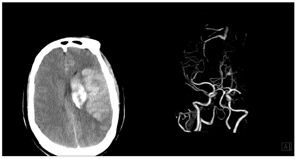
[0037] At 12:20 a.m. on March 20, 2015, the patient, female, 77 years old, was sent to Chongqing Southwest Hospital due to sudden right limb weakness for 2 hours. The muscle strength of the right limb was grade 0, lethargy, and the body temperature was 36.2 ℃. Using the infrared thermal imager described in Example 1, the patient's right temporal, right forehead, eyebrow center, left forehead, and left temporal were quickly measured at five points simultaneously, and the temperature measurement and recording process was completed at 12:25 p.m. see Figure 5 . It can be seen that the temperature of the left frontotemporal region is significantly higher than that of the right frontotemporal region, and the temperatures at five points are all higher than 36.5°C.
[0038] Then, at 12:55 p.m., the head CT examination was completed. The plain CT scan showed cerebral hemorrhage in the left basal ganglia area, pressure on the left lateral ventricle, and hemorrhage, and the bleeding v...
PUM
 Abstract
Description
Claims
Application Information
 - R&D
- Intellectual Property
- Life Sciences
- Materials
- Tech Scout
- Unparalleled Data Quality
- Higher Quality Content
- 60% Fewer Hallucinations
Browse by: Latest US Patents, China's latest patents, Technical Efficacy Thesaurus, Application Domain, Technology Topic, Popular Technical Reports.
© 2025 PatSnap. All rights reserved.Legal|Privacy policy|Modern Slavery Act Transparency Statement|Sitemap|About US| Contact US: help@patsnap.com


