Construction method and device of three-dimensional model, image monitoring method and device

A three-dimensional model and construction method technology, applied in the medical field, can solve the problems of lack of tissue texture information, inability to adapt to clinical practice, inability to provide patient-specific feature information, etc., to achieve the effect of improving accuracy

Inactive Publication Date: 2015-11-25
CHONGQING HAIFU (HIFU) TECHNOLOGY CO LTD
View PDF11 Cites 27 Cited by
  • Summary
  • Abstract
  • Description
  • Claims
  • Application Information

AI Technical Summary

Problems solved by technology

Some scholars propose to use digital human body to guide ultrasonic monitoring. However, the data source of digital human body is generally to use digital camera to slice and shoot the corpse model. The whole human body model can be established by three-dimensional reconstruction in the computer. If you want to establish For the tissue anatomical model of a specific part, different tissues need to be manually or automatically outlined, and then color-matched for different tissues. After three-dimensional reconstruction, the tissue anatomical model can be obtained, and the positional relationship between different tissues can be adjusted separately. But the model generally does not have tissue texture information
At the same time, the digital human body can only be used as a common model, and cannot provide patient-specific characteristic information
Therefore, the use of digital human body to guide ultrasound monitoring of HIFU non-invasive treatment and needle minimally invasive treatment has limitations that cannot be adapted to clinical practice.

Method used


Image

  • Construction method and device of three-dimensional model, image monitoring method and device
  • Construction method and device of three-dimensional model, image monitoring method and device
  • Construction method and device of three-dimensional model, image monitoring method and device

Examples


Embodiment 1

[0071] This embodiment provides a method for building a three-dimensional model, including the following steps:

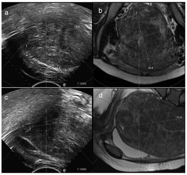
[0072] Step 1: Obtain in real time the monitoring image of the tissue in the monitoring area during the treatment of the patient, the monitoring image is a two-dimensional ultrasound image, and obtain the tissue in the monitoring image (two-dimensional ultrasound image) from the patient's three-dimensional anatomical model Diagnostic images with similar feature information, the diagnostic images are two-dimensional ultrasound / MR / CT images;

[0073] Step 2: Registering the monitoring image (two-dimensional ultrasound image) with the diagnostic image (two-dimensional ultrasound / MR / CT image) used to construct the three-dimensional model to obtain a two-dimensional registration image;

[0074] Step 3: Construct a 3D model according to the 2D registered images.

[0075] Wherein, the feature information similarity of the tissues mainly refers to the similarity in the si...

Embodiment 2

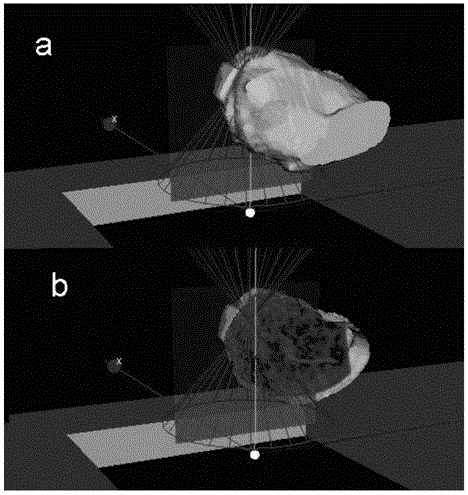
[0079] like figure 1 As shown, this embodiment provides a method for constructing a three-dimensional model, including the following steps:

[0080] Step 1: Obtain in real time the monitoring image of the tissue in the monitoring area during the treatment of the patient, the monitoring image is a two-dimensional ultrasound image, and obtain a diagnostic image similar to the characteristic information of the tissue in the monitoring image from the patient's anatomical model , the diagnostic image is a two-dimensional ultrasound / MR / CT image.

[0081] In this embodiment, the ultrasonic probe is used to obtain real-time monitoring images of tissues in the monitoring area during treatment of patients.

[0082] Among them, the anatomical model used in this embodiment is established through the patient's recent diagnostic images (such as two or three days before treatment), so it is basically consistent with the patient's tissue lesion information during treatment, but due to the T...

Embodiment 3

[0119] This embodiment provides an image monitoring method, including the following steps:

[0120] Step 1: acquiring or constructing the three-dimensional model obtained in Example 2;

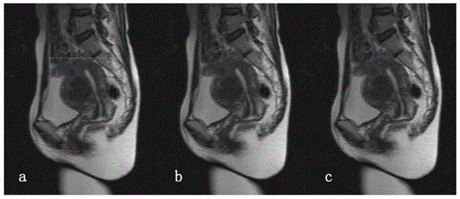
[0121] Step 2: According to the monitoring image of the tissue in the monitoring area acquired in real time during the treatment of the patient, a diagnostic image consistent with the characteristic information of the tissue in the monitoring image is obtained in the three-dimensional model, and the diagnostic image is a two-dimensional cutting image. The two-dimensional cutting image is used as a navigation image for guiding treatment.

[0122] The two-dimensional cutting image is specifically a two-dimensional cutting image that is displayed in a specific window by cutting tissue texture information along a virtual ultrasound scanning plane in the three-dimensional model according to the monitoring image.

[0123] Preferably, the image monitoring method may further include the following ste...


PUM


Abstract

A three-dimensional model construction method and device, and an image monitoring method and device. The three-dimensional model construction method comprises the steps of: 1) acquiring a monitoring image (a real-time two-dimensional ultrasonic image during treatment) of a tissue within a monitoring region during the treatment of a patient in real time, and acquiring a diagnosis image (a two-dimensional ultrasonic / MR / CT image acquired before treatment) similar to feature information about a monitoring image tissue from an anatomical model of the patient; 2) registering the monitoring image and the diagnosis image, and establishing a registration relationship to obtain a two-dimensional registration image; and 3) constructing a registered three-dimensional model according to the two-dimensional registration image. The constructed three-dimensional model can realize relatively high registration with the monitoring image during the treatment of the patient, and display clear tissue boundary and tissue texture information at the same time, so as to assist a doctor to conduct ultrasonic locating and monitoring.

Description

technical field [0001] The invention belongs to the field of medical technology, and relates to a three-dimensional model construction method, a three-dimensional model construction device, an image monitoring method and an image monitoring device. Background technique [0002] In the process of MR / CT-guided ultrasound-monitored HIFU noninvasive treatment and needle minimally invasive treatment, doctors use ultrasound to locate and monitor the position of the target area. However, ultrasound images (two-dimensional images) are characterized by obvious tissue boundaries, while tissue texture Very vague, MR / CT-guided ultrasound monitoring is used for 3D reconstruction of the patient’s ultrasound / MR / CT images, and realizes the 3D registration of the 3D model and the patient’s body position during treatment according to various registration techniques, and then obtains the ultrasound scanning surface The corresponding MR / CT cutting images provide doctors with clear tissue textur...

Claims


Application Information

Patent Timeline
Patent Type & Authority Applications(China)
IPC IPC(8): A61B8/08A61B5/055A61B6/03G06T17/00G06T7/00
CPCA61B8/08
Inventor 文银刚
Owner CHONGQING HAIFU (HIFU) TECHNOLOGY CO LTD
Why Eureka
Try Eureka