
Automatic focusing method and device used for microscope

An automatic focusing and microscope technology, applied in the field of microscopes, can solve the problem of not being suitable for high-magnification microscopic imaging systems

Inactive Publication Date: 2015-11-25
SIEMENS HEALTHCARE DIAGNOSTICS INC
View PDF8 Cites 13 Cited by
  • Summary
  • Abstract
  • Description
  • Claims
  • Application Information

AI Technical Summary

Problems solved by technology

However, this method is not suitable for the high-magnification microscopy imaging systems used in urine sediment analysis

Method used


Image

  • Automatic focusing method and device used for microscope
  • Automatic focusing method and device used for microscope
  • Automatic focusing method and device used for microscope

Examples


Embodiment Construction

[0030] In order to make the object, technical solution and advantages of the present invention clearer, the present invention will be further described in detail below with reference to the accompanying drawings and examples.

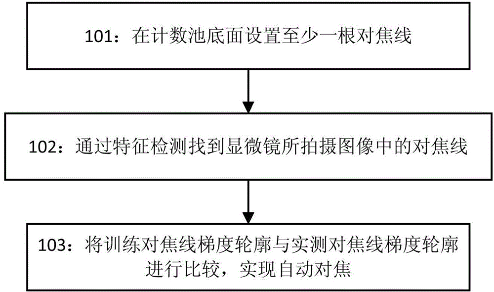
[0031] Embodiments of the present invention provide an automatic focusing method and a microscopic imaging system, which can realize real-time focusing. figure 1 It is a schematic flowchart of an autofocus method for a microscope in an embodiment of the present invention, including the following steps.

[0032] Step 101: Pre-set at least one focus line on the bottom surface of the counting tank.

[0033] For the microscope slide used for cell counting, there is a groove in the center of the slide, which serves as a counting chamber, and its depth direction is the z-axis direction. In one embodiment of the present invention, grid-shaped focal lines can be marked on the bottom surface of the groove (x-y plane). In a specific implementation, when setting...


PUM


Abstract

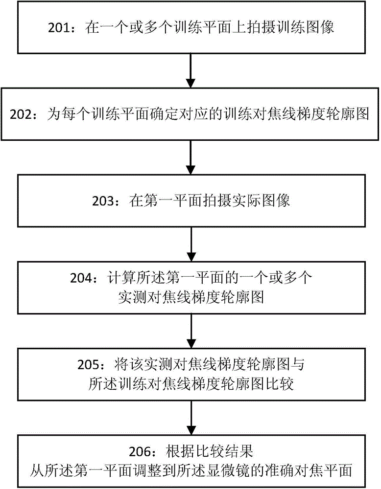
The embodiment of the present invention discloses an automatic focusing method and device used for a microscope. The method comprises the steps of adjusting the microscope on a or a plurality of training planes to shoot a training image containing at least one focusing line, finding out the focusing line from the image, and determining a corresponding training focusing line gradient profile diagram for each training plane; arranging the at least one focusing line on the bottom surface of a counting pool; adjusting the microscope to a first plane to shoot an actual image, finding out the focusing line from the image, and calculating a or a plurality of actual measurement focusing line gradient profile diagrams of the first plane; determining a training focusing line gradient profile diagram matching the one or the plurality of actual measurement focusing line gradient profile diagrams, and adjusting the microscope from the first plane to an accurate focusing plane according to the relative position relation of a second plane corresponding to the matched training focusing line gradient profile diagram and the accurate focusing plane.

Description

technical field [0001] The invention relates to the technical field of microscopes, in particular to an automatic focusing method and device for microscopes. Background technique [0002] Before applying image processing and pattern recognition techniques to microscopic images of cells and other tiny particles, such as those formed from urine, researchers need to obtain images that are precisely in focus. Using an autofocus system for image capture saves time and provides better and more consistent focus than humans taking pictures. With the introduction of fast automatic analysis instruments such as urine sediment analyzers in the fields of biology and medicine, high-speed microscopes are becoming more and more important. A critical step in the high-speed automated microscope image capture process is instantaneous focusing. Fast and reliable autofocus methods are essential for large-scale microscopy image acquisition, capture. [0003] In traditional technology, for auto...

Claims


Application Information

Patent Timeline
IPC IPC(8): G02B7/28G02B21/36
Inventor 迟颖沈宏
Owner SIEMENS HEALTHCARE DIAGNOSTICS INC
Learn More