Medical image diagnostic apparatus and medical image processing apparatus

A technology for medical images and diagnostic devices, applied in image data processing, instruments for radiological diagnosis, diagnosis, etc., can solve problems such as complicated work, and achieve the effect of reducing work and simple operation

Active Publication Date: 2016-01-27
TOSHIBA MEDICAL SYST CORP
View PDF18 Cites 4 Cited by
  • Summary
  • Abstract
  • Description
  • Claims
  • Application Information

AI Technical Summary

Problems solved by technology

[0006] However, since these series of operations are required for the inspection, the work during the inspection becomes cumbersome

Method used


Image

  • Medical image diagnostic apparatus and medical image processing apparatus
  • Medical image diagnostic apparatus and medical image processing apparatus
  • Medical image diagnostic apparatus and medical image processing apparatus

Examples


no. 1 approach

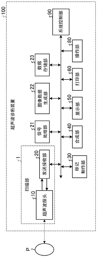
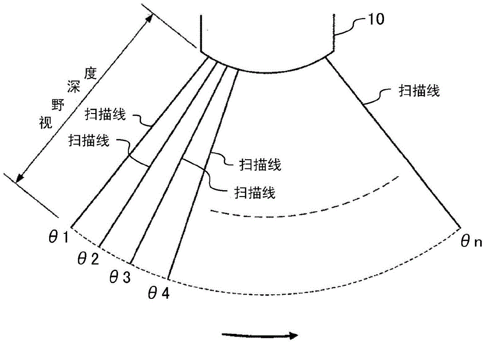
[0033] In the first embodiment, an ultrasonic diagnostic apparatus will be described as an example of a medical image diagnostic apparatus. figure 1 It is a block diagram showing the configuration of the ultrasonic diagnostic apparatus according to the first embodiment. This ultrasonic diagnostic apparatus 100 includes a scanning unit 1 . The scanning unit 1 executes scanning for generating images of the inside of the subject P. As shown in FIG. For example, the scanning unit 1 includes: an ultrasonic probe 10 for scanning the subject P by ultrasonic waves; deal with. In addition, a signal processing unit 21 that generates data based on the signal processed by the transmitting and receiving unit 20 , and an image data generating unit 22 that generates image data based on the data generated by the signal processing unit 21 are provided.

[0034] In addition, the ultrasonic diagnostic apparatus 100 includes: a data storage unit 23 storing the image data generated by the image...

no. 2 approach

[0135] In the first embodiment, a case was described in which the medical imaging diagnostic apparatus is an ultrasonic diagnostic apparatus, but the embodiment is not limited thereto. For example, an MRI (Magnetic Resonance Imaging, Magnetic Resonance Imaging) device is sometimes used to measure fetal development. In such a case, in order to reduce the work in inspection by simple operation, it is also possible to apply a mark generation process. Therefore, in the second embodiment, a marker generation process in a medical imaging diagnostic apparatus other than an ultrasonic diagnostic apparatus will be described.

[0136] In addition, the configuration example of the medical imaging diagnostic apparatus according to the second embodiment is different from that of figure 1 The configuration example of the ultrasonic diagnostic apparatus 100 shown is the same. That is, the medical image diagnostic apparatus according to the second embodiment includes a scanner unit 1 , a si...


PUM


Abstract

The embodiment method relates to a medical image diagnostic apparatus and a medical image processing apparatus. A medical image diagnostic apparatus and a medical image processing apparatus which can reduce working in checking through simple operation. A medical image diagnostic apparatus according to the embodiment includes a scanner, image generating circuitry, marker generating circuitry, and control circuitry. The scanner performs scanning to generate an image of the inside of a subject. The image generating circuitry generates an image based on the result of scanning performed by the scanner. The marker generating circuitry generates a marker provided with information at a position serving as a reference for comparison with a certain structure. The control circuitry displays the image and the marker on the same screen of a display.

Description

[0001] Citations from related applications: [0002] This application enjoys the benefit of priority of Japanese Patent Application No. 2014-148175 filed on Jul. 18, 2014, the entire contents of which are incorporated herein by reference. technical field [0003] Embodiments relate to a medical image diagnosis device and a medical image processing device. Background technique [0004] An ultrasonic diagnostic apparatus transmits ultrasonic waves into the subject and receives ultrasonic waves reflected according to the difference in acoustic impedance of the subject tissue through a simple operation of bringing an ultrasonic probe into contact with the body surface of the subject. Furthermore, image data can be generated based on the received ultrasonic waves, and the generated image data can be displayed on the monitor in real time. Therefore, it can be widely used in examinations of the heart, blood vessels, abdomen, urinary organs, obstetrics, and gynecology in vivo. [...

Claims


Application Information

Patent Timeline
Patent Type & Authority Applications(China)
IPC IPC(8): A61B8/00G06T7/00G06T11/00
CPCA61B6/032A61B8/085A61B8/468A61B8/0866A61B8/463A61B8/14A61B6/468A61B5/055A61B2503/02A61B5/4362
Inventor 栗田康一郎渡边正毅后藤英二福田省吾
Owner TOSHIBA MEDICAL SYST CORP
Why Eureka
Try Eureka