Method and apparatus for segmenting three-dimensional medical image

A medical image, three-dimensional technology, applied in image enhancement, image analysis, image data processing and other directions, can solve the problem that segmentation method cannot accurately locate and segment polyp tissue, and it is easy to miss wide-based and flat polyps.

Inactive Publication Date: 2016-02-24
NEUSOFT MEDICAL SYST CO LTD
View PDF0 Cites 16 Cited by
  • Summary
  • Abstract
  • Description
  • Claims
  • Application Information

AI Technical Summary

Problems solved by technology
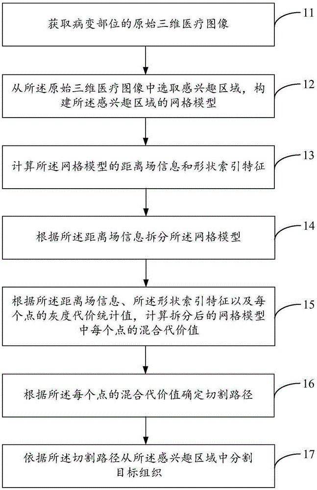
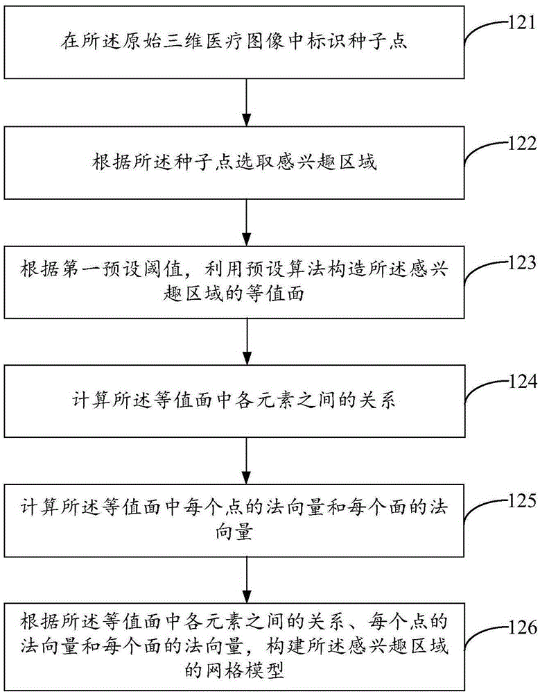
However, actual colonic polyps can be divided into: broad-based type, pedunculated type, and flat type according to the surface type. The segmentation method provided by related technologies is only applicable to pedunculated polyps, and it is easy to miss broad-based type and flat typ...

Method used


Image

  • Method and apparatus for segmenting three-dimensional medical image
  • Method and apparatus for segmenting three-dimensional medical image
  • Method and apparatus for segmenting three-dimensional medical image

Examples


Embodiment Construction

[0110] The terminology used in the present invention is for the purpose of describing particular embodiments only and is not intended to limit the invention. As used herein and in the appended claims, the singular forms "a", "the", and "the" are intended to include the plural forms as well, unless the context clearly dictates otherwise. It should also be understood that the term "and / or" as used herein refers to and includes any and all possible combinations of one or more of the associated listed items.

[0111] In the process of medical imaging, three-dimensional image data representing one or more physical properties inside human tissue can be obtained through a variety of non-invasive inspection methods. Three-dimensional medical images, such as CT (Computed Tomography, electronic computer tomography), PET (Positron Emission Tomography, positron emission computer tomography), MRI (Magnetic Resonance Imaging, magnetic resonance imaging), ultrasound and other medical imaging...


PUM


Abstract

The present application provides a method and an apparatus for segmenting a three-dimensional medical image. The method comprises: acquiring an original three-dimensional medical image of a lesion site; from the original three-dimensional medical image, selecting a region of interest so as to construct a mesh model of the region of interest; calculating distance field information and a shape index feature of the mesh model; segmenting the mesh model according to the distance field information; calculating a mixture cost value of each point in a segmented mesh model according to the distance field information, the shape index feature and a gray cost statistical value of each point; determining a segmentation path according to the mixture cost value of each point; and segmenting target tissue from the region of interest according to the segmentation path. With adoption of the method for segmenting a three-dimensional medical image provided by the present disclosure, diseased tissue in the original three-dimensional medical image can be accurately positioned and segmented, thereby improving the accuracy of three-dimensional medical image segmentation.

Description

technical field [0001] The present application relates to the technical field of medical image segmentation, in particular to a method and device for segmenting three-dimensional medical images. Background technique [0002] Medical image segmentation is the basis for subsequent processing such as 3D visualization of normal and diseased tissues, surgical simulation, and graphic-guided surgery. The accuracy of segmentation is crucial for doctors to judge the true condition of a patient's disease and make a corresponding diagnosis plan. [0003] Taking the segmentation of a CT image of colon polyps as an example, in the related art, the method of segmenting colon polyp images is based on the spherical or hemispherical geometric structure of the diseased tissue, and depends on parameters such as shape curvature to determine the segmentation plane of colon polyps. However, actual colonic polyps can be divided into: broad-based type, pedunculated type, and flat type according to ...

Claims


Application Information

Patent Timeline
IPC IPC(8): G06T7/00
CPCG06T17/30G06T2207/10081G06T2207/10088G06T2207/10104G06T2207/10136G06T2207/30032
Inventor 李丙生何薇
Owner NEUSOFT MEDICAL SYST CO LTD
Why Eureka
Try Eureka