Portal imaging for brachytherapy

A technology of imaging and radiation sources, applied in the field of therapy, can solve the problems of not being able to distinguish radioactive seeds, etc.

Inactive Publication Date: 2016-03-16
KONINKLJIJKE PHILIPS NV
View PDF3 Cites 1 Cited by
  • Summary
  • Abstract
  • Description
  • Claims
  • Application Information

AI Technical Summary

Problems solved by technology

Ultrasound guidance allows assessment of needle location but cannot resolve radioactive seeds throughout the prostate volume

Method used


Image

  • Portal imaging for brachytherapy
  • Portal imaging for brachytherapy

Examples


Embodiment Construction

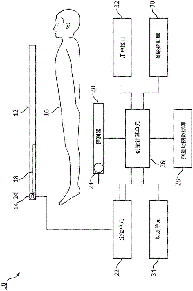
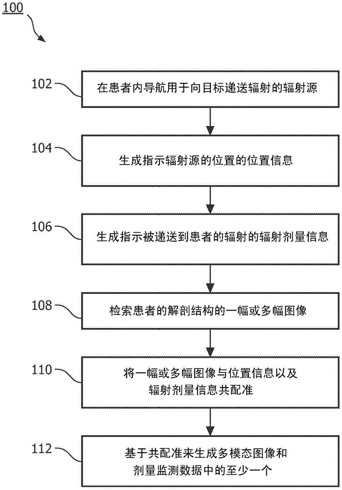
[0017] This application relates to a real-time localization and dose monitoring system for radiographic imaging for brachytherapy. Specifically, a real-time, high-accuracy positioning system is utilized to track a moving brachytherapy source, and in some embodiments, radiation detector(s). The localization system can be an optical shape sensing system (OSS) that tracks fiber sensors integrated into the source and detector(s). Positioning information from other means such as differential GPS (dGPS), impedance sensing, optical marker / camera measurements, or electromagnetic (EM) tracking is also contemplated. Information gathered by detection of emitted radiation and localization systems is combined with other imaging information derived from, for example, (TR)US, fluoroscopy, CT or optical, MRI, fluoroscopic or infrared imaging to provide information including patient and organ Motion / deformed multimodal images or dose monitoring data for real-time dose verification and adaptiv...


PUM


Abstract

A system for interventional brachytherapy for generating data to be used directly for therapy and / or for therapy planning includes a radiation source which irradiates tissue of a patient and one or more radiation detectors which detect radiation delivered to the patient and generate radiation dosage data indicative thereof. One or more position sensors determine the position of the radiation source and a localization unit, in communication with the one or more position sensors, generates position data indicative of the position of the radiation source. An image database stores one or more anatomical images of the patient. A dose calculation unit which co-registers the one or more anatomical images with the positional and radiation dosage data and generates dose monitoring data based on the co-registration

Description

technical field [0001] This application relates to therapeutic techniques. This application finds particular application in connection with, and will be described with particular reference to, high dose rate (HDR) brachytherapy. However, it is to be appreciated that the present invention will also be used in connection with other therapeutic treatments such as low dose brachytherapy, localizing other treatment sources, and the like. Background technique [0002] The more focal radiation therapy (RT) develops, the more important dose monitoring becomes in order to track and adapt dose to target volumes or critical structures. Portal imaging is the acquisition of images using radiation therapy beams. Images from these devices are then used, for example, to verify the patient's treatment. Absorbed dose, patient positioning, organ motion / deformation are monitored in situ during external beam RT in orthogonal radiography and in-beam PET. During portal imaging, target volume a...

Claims


Application Information

Patent Timeline
Patent Type & Authority Applications(China)
IPC IPC(8): A61N5/10
CPCA61N5/1001A61N5/1049A61N5/1064A61N2005/1022A61N2005/1058A61N5/1071A61B2034/2051A61B2034/2061A61N5/1039
Inventor C·里宾R·陈L·F·古铁雷斯
Owner KONINKLJIJKE PHILIPS NV
Why Eureka
Try Eureka