
Method of image guided intraoperative simultaneous several ports microbeam radiation therapy with microfocus X-ray tubes

a microfocus x-ray tube and imaging guided technology, applied in the field of image guided intraoperative simultaneous several ports microfocus radiation therapy with microfocus x-ray tube, can solve the problems of not being able to achieve synchrotron radiation dose rate, not being able to achieve a present medical accelerator or an orthovoltage tube, and not being able to achieve a present medical accelerator

Inactive Publication Date: 2017-05-02
SAHADEVAN VELAYUDHAN
View PDF37 Cites 40 Cited by
  • Summary
  • Abstract
  • Description
  • Claims
  • Application Information

AI Technical Summary

Benefits of technology

This patent describes a new and advanced radiation therapy method using multiple simultaneous X-ray microbeams. The treatment involves using multiple microfocus X-ray tubes that can be placed close to the tumor or organ to be treated. The microbeams have a high dose rate, which is important for effective treatment. The therapy can be performed using external or interstitial implants, and can also be combined with other treatments like electronic brachytherapy or carbon nanotube field emission X-ray tubes. The therapy can be used for a variety of cancer treatments and also has applications in regenerating acutely injured or chronically diseased organs. The invention offers a low-cost, high-quality radiation therapy machine that is affordable and minimally invasive.

Problems solved by technology

However, its dose rate is nowhere near to that of synchrotron radiation.
It is not achievable with a present medical accelerator or from an orthovoltage tube.
It is not achievable with a present medical accelerator.
Hence the increased spacing of the microbeam do not minimize dose to the tumor.
Such high dose and dose rate radiation therapy is not possible with present medical accelerators.
Its radiobiological effectiveness for tumor cure and control is much inferior as compared to that of synchrotron radiation.
It definitely cannot provide anywhere closer to the radiobiological effectiveness of synchrotron radiation with dose rate of 20,000 Gy / sec in treating a tumor.
The other major disadvantage of the X-ray tube based microbeam radiation as taught in U.S. Pat. No. 7,643,610 is its 0.8 mm sized large penumbra (18).
Using the 1 mm wide microbeam as in U.S. Pat. No. 7,643,610 (19) is a significant hazard as it increases the normal tissue damage and necrosis.
However, synchrotrons are not readily available to most of the cancer treatment centers in the world.
Hence its inferior RBE and poor tumor cure and control.
It definitely cannot provide anywhere close to the radiobiological effectiveness of synchrotron radiation.
It results in tumor recurrence.
However, it does not represent the additive isodose of simultaneous beams.
To treat a second patient, a second source is used, hence it is relatively expensive to maintain.
Because of the potential high dose to the skin, to the chest wall and the ribs for patients with small breast, this system cannot be used for accelerated partial breast irradiation (APBI) for such patients.
Even for those patients who are eligible for APBI with this system, there is the potential danger of balloon deflation and deformity.
It can cause skin fibrosis, rib fracture and breast deformity.
Furthermore, its voltage is more easily controlled and it is very compact.
This causes another week long waiting period for this third surgery.
If additional boost radiation to the tumor bed is also given it will also take additional time.
Because of the poor dose rate of the orthovoltage X-ray beam, 0.17 to 0.83 Gy / sec, it does not offer the same radiobiological advantages of stereotactic microbeam radiosurgery with synchrotron radiation with the dose rate of 20,000 Gy / sec.
The tumor recurrence is primarily due to conventional radiation therapy's inability to sterilize cancer stem cells.
The “differentiated” cancer cells might be sterilized with conventional radiation therapy but the residual radioresistant cancer stem cells could cause tumor recurrence.
The low dose rate conventional radiation therapy is less effective for sterilization of the cancer stem cell.
However when it is compared with excision alone, it has no benefit in improving survival or reducing distant metastasis (73).
However, in general community practice, it is seldom one sees a patient who was treated by breast preservation surgery and whole breast radiation with or without boost radiation to the tumor bed without a great deal of breast deformity.
Most often, its cosmetic outcome is very poor.
It is in addition to the economical loss from time taken away from work to attend to the treatments.
Thus when radiation therapy is rendered with parallel microbeam with broad beam effect at the tumor site, the tolerance dose to the normal issue is extremely high.
It is the reason why it does not visualize the soft tissue adequately.
The 10 keV photons will not penetrate the skin sufficiently and hence it is not used to treat deep seated tumors.
However, without any added applicator protective cover, this 10 kV X-ray is locally absorbed.
Hence its RBE is also very poor.
However estrogen receptor negative tumors may not have the ability to transport estrogen into the cell interior hence its poor metabolic utilization.
On a global basis, very few can afford the present IMRT or even a simple accelerator based radiation therapy system in lieu of now obsolete cobalt machines.
Many global medical institutions have advanced surgical capabilities but cannot afford the investment for the present-day medical accelerators and its accessories and needed personnel to operate them.
Present-day cancer drugs are very expensive even for economically advanced countries.
They are unaffordable to most patients from economically disadvantaged countries.
To keep a patient as immobilized and without physiologic organ movement for 50 minutes is not an achievable goal even if the patient is kept under anesthesia.
It also suffers the difficulties associated with prolonged treatment time.
Unfortunately, it is compared with radiobiologically very ineffective 0.17 to 0.83 Gy / sec X-ray tube based radiation in U.S. Pat. No. 7,643,610—by Dilimanian (87).
Abnormal activity in these subthalamic nucleuses will cause abnormal stimulus in thalamus which exerts control over the motor cortex resulting in suppression of thalamocortical activity which leads to the expression of Parkinson's disease.
The methods of microbeam radiation therapy with external 100-250 keV X-rays as in U.S. Pat. No. 7,194,063 (68) and U.S. Pat. No. 7,746,979 (55) by Dilmanian et al is difficult to implement.
However, the absorption radiology imaging systems are incapable of soft tissue imaging; they are incapable of gathering detailed information on the tumor tissue since most of the tumor is made of soft tissue.
Obviously, it is not very accurate.

Method used


Image

  • Method of image guided intraoperative simultaneous several ports microbeam radiation therapy with microfocus X-ray tubes
  • Method of image guided intraoperative simultaneous several ports microbeam radiation therapy with microfocus X-ray tubes
  • Method of image guided intraoperative simultaneous several ports microbeam radiation therapy with microfocus X-ray tubes

Examples


Embodiment Construction

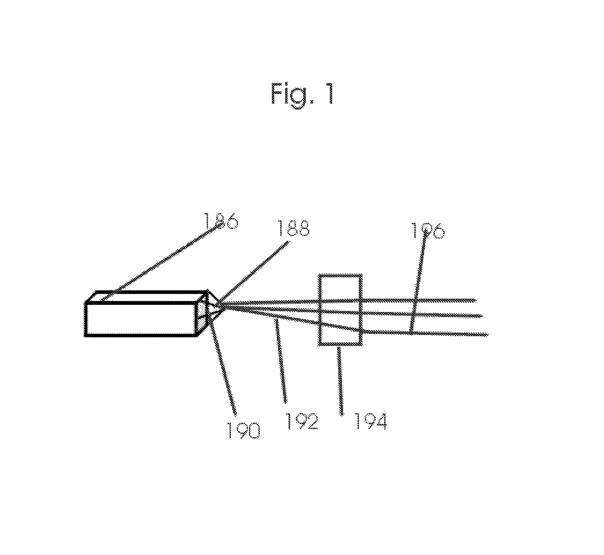
[0244]FIG. 1 illustrates a commercially available micro focus x-ray tube 186 that is equipped with analyzer crystal 190 that filters the bremsstrahlung polychromatic x-ray beam 188 to mostly monochromatic. These commercially available x-ray tubes are equipped with adjustable focal spots ranging from as low as 5 to 50 μm. The 5 μm sized focal spot is very close to the laser produced x-ray's focal spot. They are available with targets ranging from chromium, copper, molybdenum, tungsten etc. The fully packaged commercial tubes have remote control and software controlled operational capabilities. They meet all the radiation safety precautions including instructions and warnings signs on safe operation that is displayed on its display panel.

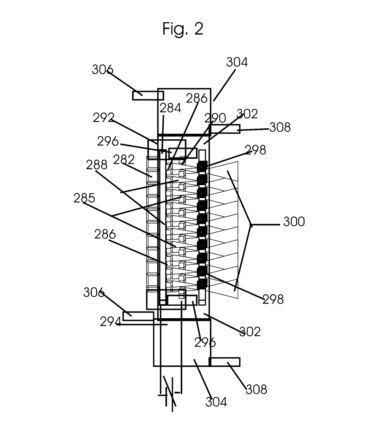
[0245]FIG. 2 is a detailed illustration of the basic structures of a CNT based single set, 10 simultaneous converging beams X-ray tube. The miniaturized tiny CNT based field emission cathode 288 is constructed with the metal-oxide-semiconductor field-...


PUM


Abstract

This invention pertains to a method of low-cost intraoperative all field simultaneous parallel microbeam single fraction few seconds duration 100 to 1,000 Gy and higher dose radiosurgery with micro-electro-mechanical systems (MEMS)-carbon nanotube based microaccelerators. It ablates cancer cells including the mesenchymal epithelial transformation associated cancer stem cells. Microbeam brachy-therapeutic radiosurgery is performed. Microaccelerators are configured for simultaneous parallel microbeam emission from varying angels to an isocentric tumor. Their additive dose rate at the isocentric tumor is in the range of 10,000 to 20,000 Gy / s. It eliminates most tumor recurrence and metastasis which enhances cancer cure rates. It also exposes cancer antigens which induces cancer immunity. Stereotactic breast core biopsy is combined with, positron emission tomography and computerized tomography and phase-contrast imaging. Parallel microbeam brachytherapy preserves normal breast appearance. Migration of normal stem cells from unirradiated valley regions heals the radiation damage to the normal tissue.

Description

[0001]The present nonprovisional application is a Continuation-In-Part of applicant's prior U.S. nonprovisional patent application entitled “Image Guided Intraoperative Simultaneous Several Ports Microbeam Radiation Therapy with Microfocus X-Ray Tubes”, Ser. No. 12 / 929,770, filed Feb. 15, 2011, which is hereby incorporated by reference in its entirety.FIELD OF INVENTION[0002]x-ray beam therapy, class 378, 424, 530FEDERALLY SPONSORED RESEARCH[0003]None;SEQUENCE LISTING[0004]Table of Contents attached1. BACKGROUND OF THE INVENTION[0005]Synchrotron high dose rate microbeams with dose rate exceeding 20 Gy / sec is curative even for the most radiation resistant tumors like the glioblastoma multiforme (Slatkin and Dilimanian). This invention is aimed at similar high dose rate radiation therapy but with much more affordable and easily manageable microbeams from microfocus X-ray tubes that are configured to provide multiple simultaneous interlaced microbeams for simultaneous several ports irr...

Claims


Application Information

Patent Timeline
IPC IPC(8): A61N5/00A61N5/10A61B10/02
CPCA61N5/1077A61N5/1001A61B10/02A61N5/1039A61N2005/1091A61N5/1027A61N5/1084A61B10/0233A61N2005/1022A61N2005/1052A61N2005/1054A61N2005/1055
Inventor SAHADEVAN, VELAYUDHAN
Owner SAHADEVAN VELAYUDHAN
Learn More