A method for automatic recognition of hemorrhage in fundus color photographic images

A fundus image and image technology, applied in the field of image processing, can solve problems such as large contrast range and uneven illumination of fundus images, and achieve reliable diagnosis and obvious bleeding areas

Active Publication Date: 2018-06-01
SHANGHAI FIRST PEOPLES HOSPITAL +1
View PDF3 Cites 1 Cited by
  • Summary
  • Abstract
  • Description
  • Claims
  • Application Information

AI Technical Summary

Problems solved by technology

[0004] Aiming at the image quality problems caused by the uneven illumination of the fundus image, the large range of contrast changes, and the structural changes in the retina before and after laser treatment in large-scale fundus image screening scenarios, the present invention proposes a hemorrhage detection method for retinal fundus images. The bleeding area is more obvious, which is convenient for doctors to diagnose fundus lesions more accurately and provide a reliable treatment plan

Method used


Image

  • A method for automatic recognition of hemorrhage in fundus color photographic images
  • A method for automatic recognition of hemorrhage in fundus color photographic images
  • A method for automatic recognition of hemorrhage in fundus color photographic images

Examples


Embodiment Construction

[0103] Below the embodiment of the present invention is described in detail, present embodiment implements on the premise of technical scheme of the present invention, has provided concrete implementation and operation process, but protection scope of the present invention is not limited to following embodiment.

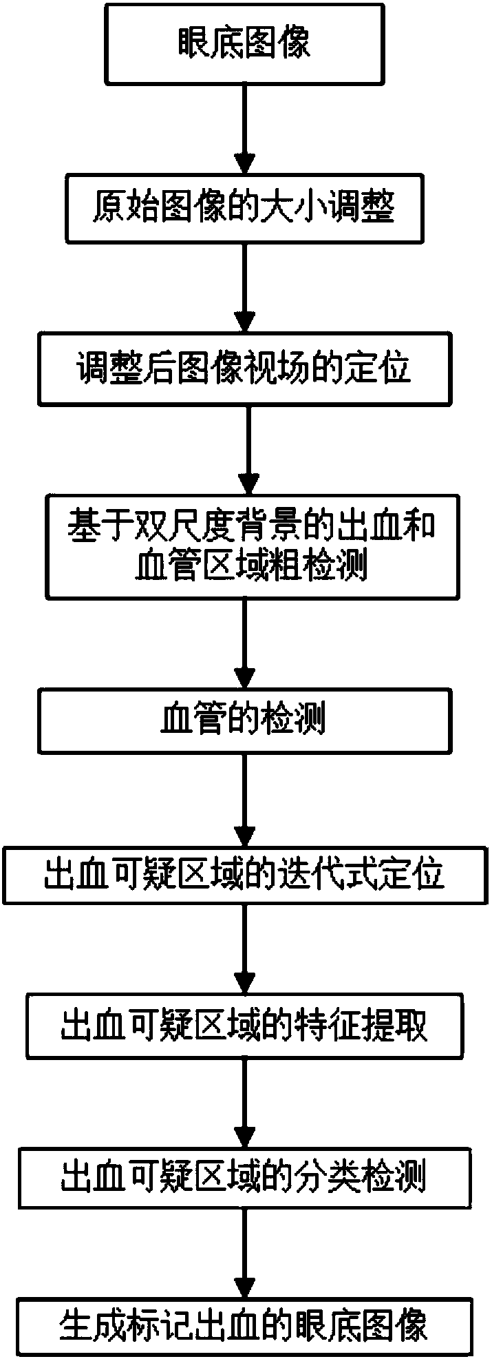
[0104] figure 1 A flow chart of the hemorrhage detection of the retinal fundus image in the embodiment of the present invention is shown. The fundus image used in this embodiment is an image taken by a color digital non-mydriatic fundus camera, such as Figure 2a Shown is the G channel component of the image.

[0105] (1) Adjust the size of the original image

[0106] In the case of large-scale fundus image screening, the size of the pictures taken by different fundus cameras may be different, so in order to make the method of the present invention applicable to the processing of images of different specifications, it is necessary to adjust the size of the original...


PUM


Abstract

The invention discloses a retinal fundus image bleeding detection method. A fundus image photographed by a color digital non-mydriatic fundus camera is utilized. The method adopts the following steps that (1) the size of an original image is adjusted; (2) the viewing field of the adjusted image is positioned; (3) bleeding and blood vessel areas based on double-scale background are crudely detected; (4) the blood vessels are detected; (5) a bleeding suspicious area is iteratively positioned; (6) feature extraction is performed on the bleeding suspicious area; and (7) classified detection is performed on the bleeding suspicious area, and the bleeding marked fundus image is generated. According to the method, the fundus image acquired under different acquisition conditions can be processed, the bleeding area can be rapidly and effectively detected and the bleeding site of the processed fundus image is visual and obvious so that further diagnosis is facilitated for ophthalmologists.

Description

technical field [0001] The invention belongs to the field of image processing, and in particular relates to a hemorrhage detection method of a retinal fundus image. Background technique [0002] Diabetic Retinopathy (Diabetic Retinopathy) is a kind of ophthalmic disease widely existing in diabetic patient, and it can have impact on patient's vision, and serious can cause the blindness of diabetic patient. Therefore, early detection and treatment of retinopathy can minimize the loss of vision. Intraretinal hemorrhage is one of the early visible signs of diabetic retinopathy. Intraretinal hemorrhages (Hemorrhages) are caused by rupture of intraretinal microaneurysms (Microaneurysms), which is an important basis for lesion staging and diagnosis. In retinal fundus images, microaneurysms appear as dark red dots, while hemorrhages appear as large dark red areas, similar in color to blood vessels, with irregular shapes and unclear borders. It is an important problem to be solved...

Claims


Application Information

Patent Timeline
Patent Type & Authority Patents(China)
IPC IPC(8): G06T7/00G06T7/11
CPCG06T7/0012G06T2207/20081G06T2207/30041
Inventor 余奇杨杰许迅周磊
Owner SHANGHAI FIRST PEOPLES HOSPITAL
Why Eureka
Try Eureka