
Cerebrovascular quantitative analysis method based on skeleton line

A quantitative analysis, skeleton line technology, applied in image analysis, medical data management, diagnostic recording/measurement, etc., can solve problems such as torsion rate not involved, and achieve an objective effect of disease diagnosis

Active Publication Date: 2016-09-07
BEIJING NORMAL UNIVERSITY
View PDF1 Cites 5 Cited by
  • Summary
  • Abstract
  • Description
  • Claims
  • Application Information

AI Technical Summary

Problems solved by technology

However, these studies on tortuosity mostly involve features such as curvature and arc length to chord ratio, and do not involve the calculation of torsion; at present, doctors only make subjective judgments on the diagnosis of diseases by looking at films, and there is no accurate data reference standard

Method used


Image

  • Cerebrovascular quantitative analysis method based on skeleton line
  • Cerebrovascular quantitative analysis method based on skeleton line
  • Cerebrovascular quantitative analysis method based on skeleton line

Examples


Embodiment Construction

[0036] The present invention will be further described below in conjunction with the accompanying drawings and embodiments.

[0037] A method for quantitative analysis of blood vessels based on skeleton lines, comprising the following steps:

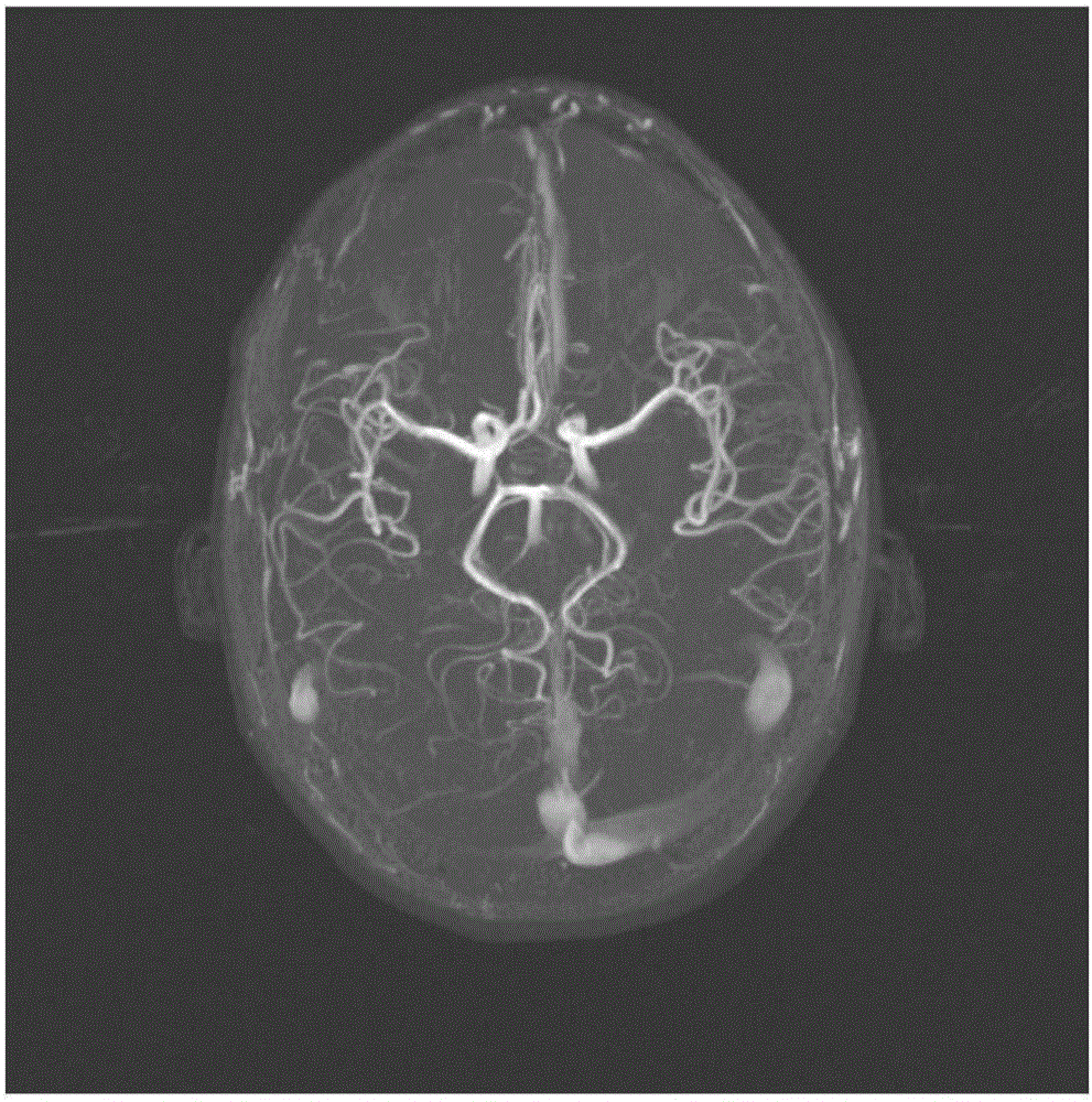
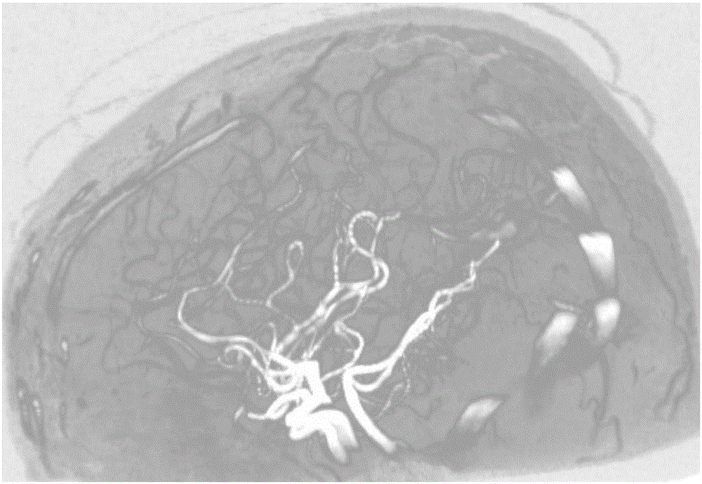
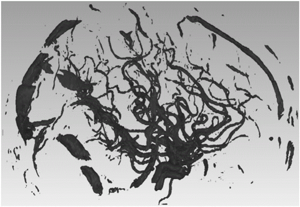
[0038] Step 1. According to the collected DICOM data of cerebral vessels, obtain the maximum density projection map of the data through perspective, and use the particle swarm optimization algorithm to segment the original data to obtain segmented volume data or Mesh data, such as figure 1 , 2 , 3 shown.

[0039] The accuracy of the segmentation results can be verified by comparing the morphological structure of the segmented data with the maximum density projection map.

[0040] Step 2: Artificially identify the part of the Wills ring from the segmented data, retain the small segment of blood vessel branches connected to the Wills ring, and delete other irrelevant blood vessels and noise. The L1-medial skeleton line extraction algori...


PUM


Abstract

The invention relates to a cerebrovascular quantitative analysis method based on a skeleton line. By use of the quantitative analysis method based on cerebrovascular DICOM data, blood vessels of a brain are segmented in original data, a Wills ring portion is identified, and a Wills ring skeleton line and a sampling point radius geometric model are obtained by performing Wills ring skeleton ring extraction by use of a skeleton line extraction algorithm. Semantic identification and rule sampling are performed on each segment of the blood vessels on a skeleton, and feature quantity information at sampling points are calculated and stored in a database. Compared to conventional two-dimensional projection image quantitative analysis and blood vessel three-dimensional volume quantitative analysis, the method provided by the invention can obtain a more accurate result. An original discrete skeleton is fitted by use of a B spline curve method, the blood vessel skeleton form is enabled to be continuous, the feature quantity of any point on the blood vessels can be obtained, and a more accurate feature quantity calculation result can be obtained.

Description

technical field [0001] The invention belongs to the field of computer graphics, and relates to the quantitative analysis of medical images in graphic geometry, in particular to a method for quantitative analysis of cerebral blood vessels based on skeleton lines. Background technique [0002] Cerebrovascular disease is a disorder in which the supply of blood vessels to the brain is dysfunctional. Severe cerebrovascular disease can lead to stroke and is likely to cause disability or death to the patient. A stroke is usually caused by a ruptured aneurysm in a blood vessel in the brain. Cerebrovascular aneurysms often occur in the circle of Wills (circle of cerebral arteries). Therefore, the research on the circle of Willis and its quantitative analysis are very important for diseases such as aneurysm rupture and cerebral infarction. The circle of Wills consists of the left and right branches of the anterior cerebral artery (ACAs), the anterior communicating artery (ACo), the ...

Claims


Application Information

Patent Timeline
Patent Type & Authority Applications(China)
IPC IPC(8): G06T7/00G06T7/60A61B5/00G06F19/00
CPCA61B5/00A61B5/489G06F19/321G06T7/0012G06T7/60G06T2207/30101G16H50/20
Inventor 王醒策刘恩惠武仲科周明全
Owner BEIJING NORMAL UNIVERSITY
Learn More