A biological tissue mass spectrometry imaging method

A mass spectrometry imaging and biological tissue technology, applied in the field of mass spectrometry imaging, can solve problems such as unfavorable optimization of mass spectrometry imaging, inability to regulate mass spectrometry signals, complex sample processing, etc., and achieve the effects of broad application prospects, high sensitivity, and accurate detection results.

Active Publication Date: 2020-10-20
SOUTH CHINA NORMAL UNIVERSITY
View PDF3 Cites 0 Cited by
  • Summary
  • Abstract
  • Description
  • Claims
  • Application Information

AI Technical Summary

Problems solved by technology

[0004] Traditional mass spectrometry imaging has the following disadvantages: (1) It is slow and cannot be directly imaged; (2) The matrix interference is serious and the sample pretreatment is complicated; (3) The ionization imaging technology after laser analysis cannot adjust the mass spectrometry signal in time and space , which is not conducive to optimizing the mass spectrometry image

Method used


Image

  • A biological tissue mass spectrometry imaging method
  • A biological tissue mass spectrometry imaging method
  • A biological tissue mass spectrometry imaging method

Examples


Embodiment 1

[0027] The biological tissue mass spectrometry imaging method provided in this embodiment includes the following steps:

[0028] (1) Select the isolated mouse kidney tissue, freeze and slice it, and obtain the mouse kidney tissue section;

[0029] (2) Select a corundum rod, attach the mouse kidney tissue section obtained in step (1) to the corundum rod, and let it dry naturally;

[0030] (3) Add the acriflavine solution dropwise on the mouse kidney tissue section, and let it stand to make it spread evenly;

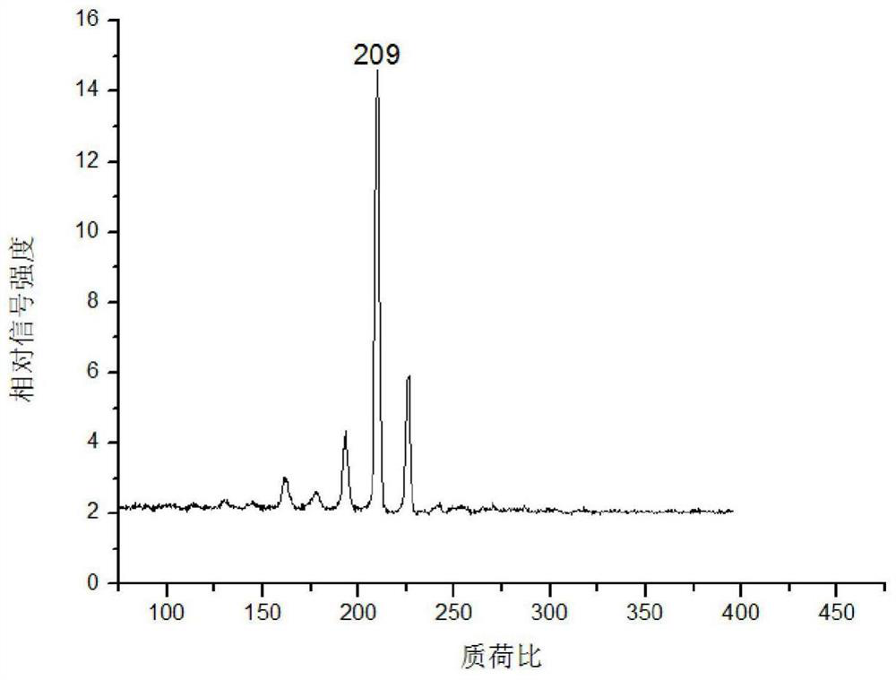
[0031] (4) Fix the corundum rod with the mouse kidney tissue slices on the injection rod of the mass spectrometer, enter the mass spectrometer with laser through the injection system for detection, and obtain the acriflavine in the mouse kidney tissue mass spectrum.

[0032] In the step (1), the mice are selected as Balb / c male mice, 7-8 weeks old.

[0033] The concentration of acriflavine solution in step (3) is 10 -3 g / mL, 10 -3 When the acriflavine solution of g / mL...

Embodiment 2

[0041] The biological tissue mass spectrometry imaging method provided in this embodiment includes the following steps:

[0042] (1) Select the isolated mouse kidney tissue, freeze and slice it, and obtain the mouse kidney tissue section;

[0043] (2) Select a corundum rod, attach the mouse kidney tissue section obtained in step (1) to the corundum rod, and let it dry naturally;

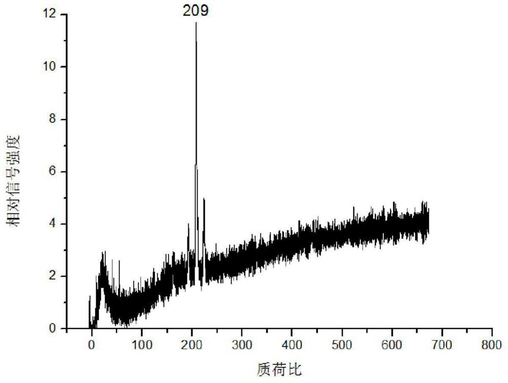
[0044] (3) Fix the corundum rod with the mouse kidney tissue slices on the injection rod of the mass spectrometer, enter the mass spectrometer with laser through the sample injection system for detection, and obtain the acriflavine in the mouse kidney tissue mass spectrum.

[0045] In the step (1), the mice are selected as Balb / c male mice, 7-8 weeks old.

[0046]In step (3), the corundum rod adhered to the mouse kidney tissue slice is fixed on the sample rod of the mass spectrometer, and then enters the mass spectrometer with laser for detection through the sample feeding system. The specific proc...

Embodiment 3

[0049] The biological tissue mass spectrometry imaging method provided in this embodiment includes the following steps:

[0050] (1) Select the isolated mouse kidney tissue, freeze and slice it, and obtain the mouse kidney tissue section;

[0051] (2) Select a corundum rod, attach the mouse kidney tissue section obtained in step (1) to the corundum rod, and let it dry naturally;

[0052] (3) 10 μL of 10 -3 g / mL acriflavine solution, let it stand to make it diffuse evenly;

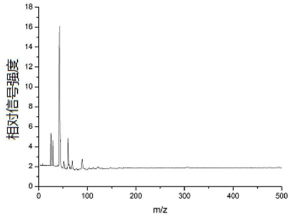
[0053] (4) Fix the corundum rod with the mouse kidney tissue slices on the injection rod of the mass spectrometer, enter the mass spectrometer with laser through the injection system for detection, and obtain the acriflavine in the mouse kidney tissue mass spectrum.

[0054] In the step (1), the mice are selected as Balb / c male mice, 7-8 weeks old.

[0055] In step (3), stand still for 15-25 minutes to make it spread evenly.

[0056] In step (4), the corundum rod adhered with the mouse kidney tissue sl...


PUM

PropertyMeasurementUnit
concentrationaaaaaaaaaa

Abstract

The invention discloses a biological tissue mass spectrometry imaging method, comprising the following steps: (1) selecting isolated mouse kidney tissue, freezing and slicing to obtain mouse kidney tissue slices; Obtained mouse kidney tissue slices adhered to the corundum rods, and air-dried naturally; (3) dripping acriflavine solution on the mouse kidney tissue slices, and let it stand to spread evenly; (4) the adhered The corundum rods of mouse kidney tissue slices dripped with acriflavine solution were placed in a mass spectrometer with a laser for detection, and the mass spectrometry data ionized after laser analysis were processed by software to obtain mouse kidneys containing acriflavine Mass spectrometry imaging of tissue. The method is fast and direct, can avoid matrix interference and has no complicated sample pretreatment process, and the ionization imaging technology after laser analysis can adjust the mass spectrometry signal in time and space, which is conducive to optimizing the mass spectrometry imaging map.

Description

technical field [0001] The invention belongs to the technical field of mass spectrometry imaging, and in particular relates to a biological tissue mass spectrometry imaging method. Background technique [0002] Laser desorption post-ionization mass spectrometry has achieved great development in the last ten or twenty years. Compared with traditional MALDI mass spectrometry, it has the advantages of being fast, direct and simple, and does not require complicated sample pretreatment. Therefore, its application is gradually emerging. In the biological field, it includes the detection of small biological molecules such as short peptides and amino acids, drug molecules, chemical additives and other chemical molecules. [0003] Mass spectrometry is a new analytical technique for studying the distribution of molecules in biological cells, tissues and living animals. It can detect and record a spatial distribution of molecules in a special system without labeling. Mass spectrometry...

Claims


Application Information

Patent Timeline
Patent Type & Authority Patents(China)
IPC IPC(8): G01N27/62
CPCG01N27/628
Inventor 胡勇军陈家新陆桥
Owner SOUTH CHINA NORMAL UNIVERSITY
Why Eureka
Try Eureka