Method for increasing definition of Micro CT of peripheral nerve image

A technology of image clarity and peripheral nerves, applied in the field of medical 3D printing biology, can solve problems such as insufficient clarity, and achieve the effect of enhancing clarity

Active Publication Date: 2016-10-12
THE FIRST AFFILIATED HOSPITAL OF SUN YAT SEN UNIV
View PDF5 Cites 0 Cited by
  • Summary
  • Abstract
  • Description
  • Claims
  • Application Information

AI Technical Summary

Problems solved by technology

Although experiments have proved that different preprocessing methods can enhance Micro CT scanning of soft tissues, there is still a problem of insufficient clarity in order to use the results of Micro CT scanning to reconstruct 3D data and perform 3D printing.

Method used


Image

  • Method for increasing definition of Micro CT of peripheral nerve image
  • Method for increasing definition of Micro CT of peripheral nerve image
  • Method for increasing definition of Micro CT of peripheral nerve image

Examples


Embodiment 1

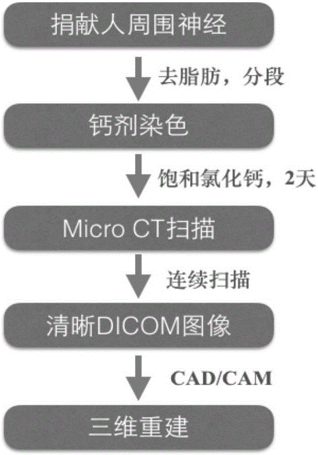
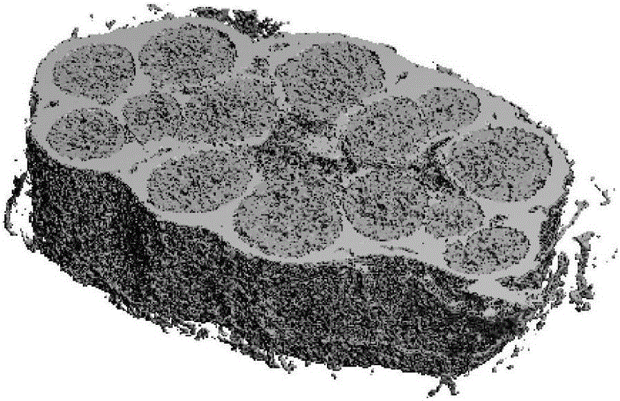
[0042] figure 1 Shown the flow chart of the method for utilizing Micro CT to scan peripheral nerve image and carry out three-dimensional reconstruction of the present invention, this method comprises the following steps:

[0043] (1) Obtain human peripheral nerves and stain them:

[0044] Remove the fat and connective tissue around the donated human peripheral nerve under a microscope, cut into small pieces; place the nerve tissue in sterile water for injection at room temperature for 7 hours; remove the sterile water for injection under aseptic operation, and soak the nerve in saturated Calcium chloride solution for 2 days, and slightly shaken; After taking out the nerve, use filter paper to absorb the surrounding water, and store it in a sealed and dry container for later use;

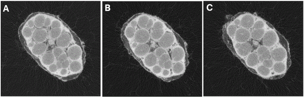
[0045] (2) Micro CT scanning of human peripheral nerves after calcium staining:

[0046] Using the nerve samples obtained in scanning step (1) of the μCT50 from SCANCO MEDICAL, Sweden, the scanning...


PUM


Abstract

The invention relates to the technical field of medical science and discloses a method for increasing definition of Micro CT of a peripheral nerve image. The method comprises the steps that soft tissues such as peripheral nerves are dyed by a solution containing calcium ions; and the Micro CT is then used for scanning of the dyed soft tissues. By the method disclosed by the invention, the definition of the scanned image reaches a requirement for nerve tract reconstruction; and the problems of insufficient accuracy and the low image definition during the peripheral nerve three-dimensional reconstruction can be solved. The method disclosed by the invention is characterized in that calcium agent dyeing is carried out to peripheral nerve samples; an original structure of the human body soft tissues is fully maintained and the structure becomes applicable to the CT; and the problem that the CT cannot be applied during the soft tissue reconstruction is solved.

Description

technical field [0001] The invention relates to a method for clearly scanning soft tissue images by using Micro CT, and belongs to the field of medical 3D printing biotechnology. Background technique [0002] With the continuous maturity of 3D printing technology, it has become possible to print tissues and organs, but soft tissues such as peripheral nerves cannot achieve true bionic printing due to the complexity of their internal structures. The reason is that it is difficult to obtain the templates required for printing. At present, in order to obtain a template for peripheral nerve printing, it is necessary to first use imaging to obtain the three-dimensional structure of the peripheral nerve, and then use computer-assisted processing of the data to generate instructions that can be used for printer operation. Therefore, it is very important to obtain clear images of peripheral nerves, which is a key step for printing. [0003] Imaging 3D reconstruction techniques mainl...

Claims


Application Information

Patent Timeline
Patent Type & Authority Applications(China)
IPC IPC(8): A61B6/00A61B6/03
CPCA61B6/03A61B6/48A61B6/50
Inventor 戚剑闫立伟刘小林朱爽林焘
Owner THE FIRST AFFILIATED HOSPITAL OF SUN YAT SEN UNIV
Why Eureka
Try Eureka