Integrated detection method for separating, enriching and detecting urine exosome as well as detection chip

An integrated detection and exosome technology, applied in the field of biotechnology detection, can solve the problems of inability to meet the detection requirements and low exosome content, and achieve the effect of easy acquisition and great application prospects

Active Publication Date: 2016-11-09
ZHEJIANG UNIV
View PDF2 Cites 15 Cited by
  • Summary
  • Abstract
  • Description
  • Claims
  • Application Information

AI Technical Summary

Problems solved by technology

However, in fact, due to the very low exosome content per unit volume of urine, the exosomes obtained by a single extraction by the general method cannot meet the requirements of subsequent detection at all. Therefore, the current detection of exosomes is mainly based on the first Concentrate the exosomes in the sample before taking certain characterization measurements

Method used


Image

  • Integrated detection method for separating, enriching and detecting urine exosome as well as detection chip
  • Integrated detection method for separating, enriching and detecting urine exosome as well as detection chip
  • Integrated detection method for separating, enriching and detecting urine exosome as well as detection chip

Examples


Embodiment 1

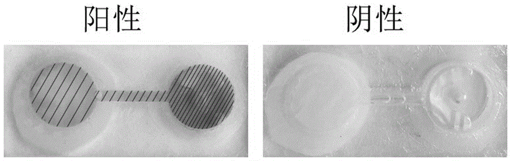
[0053] Example 1: Separation of exosomes in urine of bladder cancer patients and detection by chip ELISA method, refer to the attached Figure 1-8 .

[0054] see Figure 1-7 , an integrated detection method for urine exosome isolation, enrichment and ELISA detection, comprising the following steps:

[0055] 1) On the premise of the patient’s knowledge and the consent of the ethics committee, 16 urine samples were collected from bladder cancer patients (clinical biopsy confirmed bladder cancer), 8 healthy donors, each 100ml, and centrifuged at 2,000g at room temperature Cells and large debris were removed, and the supernatant was obtained.

[0056] 2) Ultracentrifuge the supernatant obtained in 1) at 100,000 g for 60 minutes at 4°C, discard the supernatant to collect the precipitate, and resuspend the exosomes with 1 mL of PBS buffer with pH 7.4 (such as figure 1 ).

[0057] 3) Inject the supernatant obtained in 1) into the double-membrane filter chip at a flow rate of 40 μ...

Embodiment 2

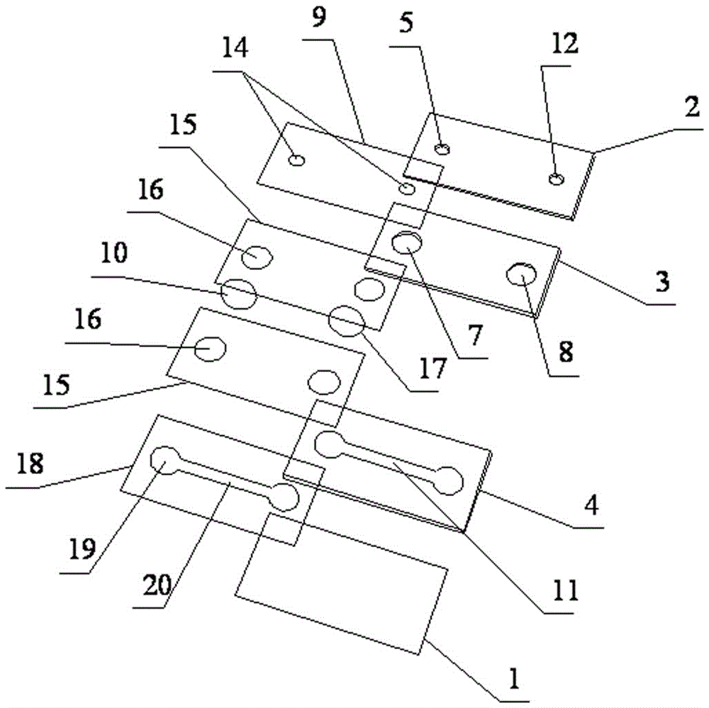
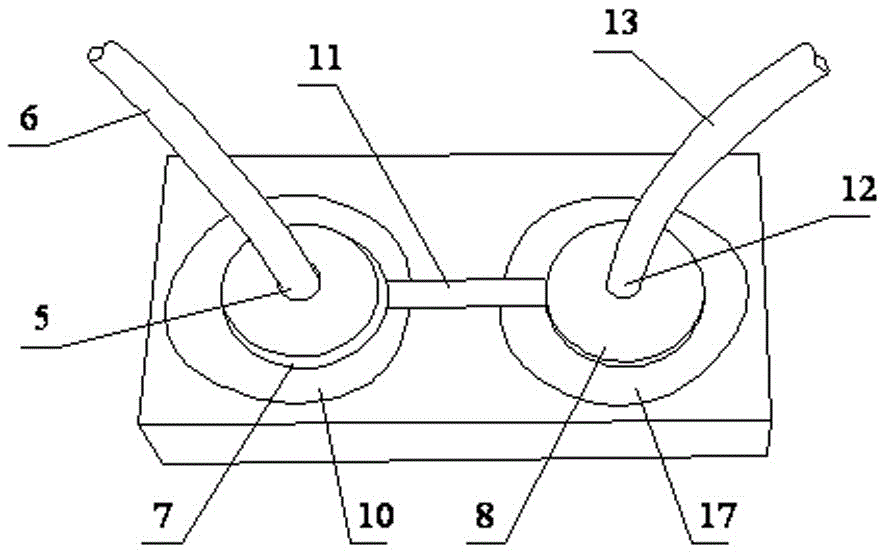
[0066] Embodiment 2, integrated detection chip, with reference to attached Figure 1-3 .

[0067] The microfluidic chip of the present invention comprises a base plate 1, a top plate 2, and a detection plate located between the base plate and the top plate, the detection plate is provided with a liquid detection cavity, and the liquid detection cavity can remove exosomes in urine It is collected in it and enriched by multiple injection samples to meet the detection requirements.

[0068] The detection board includes an upper detection board 3 and a lower detection board 4. The top board 2 is provided with a liquid injection hole 5 and a liquid discharge hole 12 formed by cutting, and the liquid injection hole 5 is connected with a liquid injection pipe 6. The liquid discharge hole 12 is connected with a liquid discharge pipe 13, and the upper layer detection plate 3 and the lower layer detection plate 4 are respectively provided with a sample injection hole 7 and a waste liqu...


PUM

PropertyMeasurementUnit
pore sizeaaaaaaaaaa
pore sizeaaaaaaaaaa
lengthaaaaaaaaaa

Abstract

The invention provides an integrated detection method for separating, enriching and detecting an exosome. The method comprises the following steps: designing and assembling a dual-membrane filtration chip, constructing an ELISA detection standard curve, collecting a sample, injecting and washing, performing chip ELISA detection, and performing data processing analysis. The invention also provides an integrated detection chip for separating, enriching and detecting a urine exosome. A reaction system and the detection method of the present invention can be used for detecting the content of a urine sample of a bladder cancer patient and the exosome in a supernatant of a bladder cancer cell line nutrient solution. The detection chip has characteristic of high specificity, generates no influence or wound on human body, and the detection method does not require the expensive precision experiment apparatus (such as an ultra centrifuge and a fluorescence microscope), and has large application prospect.

Description

technical field [0001] The invention belongs to the field of biotechnology detection, and specifically relates to the development of a microfluidic chip integrating urine exosome separation, enrichment and ELISA detection. Background technique [0002] Bladder cancer refers to malignant tumors that occur on the bladder mucosa. It is the most common malignant tumor of the urinary system and one of the ten most common tumors in the whole body. It accounts for the first place in the incidence of urogenital tumors in my country, and ranks second in the West after prostate cancer. Examination methods include routine urine examination, urine exfoliation cytology, urinary tumor markers, abdominal and pelvic B-ultrasound and other examinations. Based on the results of the above examinations, it is decided whether to perform cystoscopy, intravenous urography, pelvic CT or / and pelvic MRI to confirm the diagnosis. Among them, cystoscopy is the most important method for diagnosing bl...

Claims


Application Information

Patent Timeline
Patent Type & Authority Applications(China)
IPC IPC(8): G01N33/574
CPCG01N33/574G01N2800/34
Inventor 梁利国孔梦奇盛叶峰王书崎
Owner ZHEJIANG UNIV
Why Eureka
Try Eureka