Method for separating exosome from human placenta-derived mesenchymal stem cell source and application thereof

A technology of stem cells and exosomes, which is applied in the field of obtaining membrane vesicles involved in cell biological activities, can solve the problems of inability to guarantee the amount of exosomes obtained, uneven quality of exosomes, and lengthy step-by-step centrifugation time. The effect of improved damage repair, shortened preparation time, and simplified operation

Pending Publication Date: 2017-01-04
章毅 +7
View PDF2 Cites 45 Cited by
  • Summary
  • Abstract
  • Description
  • Claims
  • Application Information

AI Technical Summary

Problems solved by technology

The exosome extraction method mainly adopts ultracentrifugation method or expensive kit column method. However, the quality of exosome obtained by ultracentrifugation method is not uniform, the amount of exosome obtained cannot be guaranteed, and the step-by-step centrifugation time is lengthy, sometimes even due to centrifugation time or centrifugation speed. And finally can not be separated, wasting time and cost
The kit-through-column method is usually only suitable for obtaining a small amount of exosome, and it is expensive

Method used


Image

  • Method for separating exosome from human placenta-derived mesenchymal stem cell source and application thereof
  • Method for separating exosome from human placenta-derived mesenchymal stem cell source and application thereof
  • Method for separating exosome from human placenta-derived mesenchymal stem cell source and application thereof

Examples


Embodiment 1

[0043] Example 1 Isolation, preparation and identification of placental mesenchymal stem cells

[0044] Neonatal placenta authorized with maternal consent was used as a source of mesenchymal stem cells.

[0045] The postpartum fresh placenta was taken under aseptic conditions, and the placenta was preserved and transported using placenta preservation solution (see Chinese invention patent No. ZL201410028687.X) and placenta collection and storage device (see Chinese utility model patent No. ZL2014203904164). Under aseptic conditions within 48 hours, the amniotic membrane was mechanically peeled off from the placenta tissue, and then the placental chorion was carefully cut to remove as much connective tissue as possible on the chorion. Soak and rinse 3 to 5 times, and cut the rinsed chorion into about 1mm with ophthalmic scissors 3 Fragments were digested by adding type II collagenase (0.1% final enzyme digestion concentration) at 37° C. for 90 min, and shaken at 200 rpm. Add ...

Embodiment 2

[0047] Example 2 Subculture of placental mesenchymal stem cells, morphological observation and culture medium collection

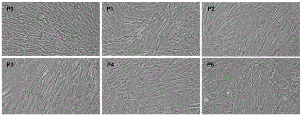
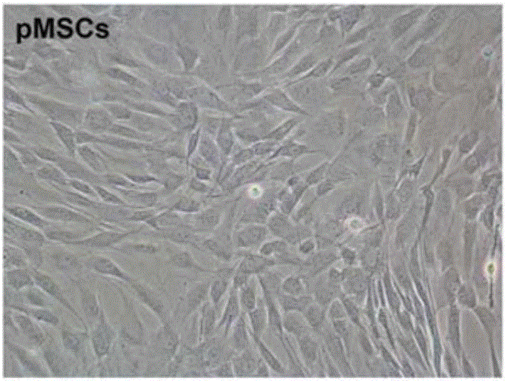
[0048] Take P0~P5 cultured cells, 5×10 4 Inoculated on 10×10 cell culture dishes, 37°C, 5% CO 2 , static culture in saturated humidity, using the serum-free medium for placental mesenchymal stem cells described in CN103805562B for culture, changing the medium every 3 days, and collecting the supernatant of the medium when the hPMSCs cell confluence reaches 85% to 90%. When the confluence of P0-P5 generation cells reached 85%-90%, the phase-contrast microscope morphology was observed and photographed (see image 3 ). Such as image 3 As shown, observed under an inverted phase-contrast microscope, 48 hours after inoculation of the primary cultured PMSCs, most of the cells adhered to the wall, and the morphology was typical fibroblast-like. Scattered some translucent round miscellaneous cells. The P0, P1, P2, and P3 generations were all long-spindle-shap...

Embodiment 3

[0049] Example 3 Acquisition of exosome

[0050] Extract 10ml-20ml of cell culture supernatant, filter it with a 0.22μm filter membrane at 4°C; then centrifuge at 10,000×g for 30min, discard the precipitate (remove subcellular components); collect the centrifuged supernatant by volume 1:1 Proportionally add 0w / v%, 9w / v%, 10w / v%, 11w / v%, 12w / v% and 13w / v% polyethylene glycol culture supernatant solution, 120,000×g for the second time After centrifugation for 75 minutes, the resulting precipitate is the exosome.

[0051] Re-suspend the precipitate with 30ml of PBS solution, mix well and then centrifuge at 120,000×g for 75min, suspend the purified exosome solution with 1ml of PBS solution, put them into eppendorf tubes, store them in a -80°C refrigerator for later use, and carry out the yield analysis. Analysis see Figure 5 , in which the 12% PEG group had the highest yield of exosome.


PUM


Abstract

The invention relates to a method for separating exosome from a human placenta-derived mesenchymal stem cell source. The method comprises the following steps: when the P2-P3 placenta-derived mesenchymal stem cell convergence rate reaches 85-90%, obtaining a cell culture supernate, carrying out membrane filtration, and collecting the filtrate; centrifugating the filtrate, collecting the centrifugal supernate, adding a 10w / v%-12w / v% polyethyleneglycol culture medium supernate solution into the centrifugal supernate according to the volume ratio of 1:1, sufficiently and uniformly mixing, and carrying out secondary centrifugation, wherein the finally obtained precipitate is the exosome. The method is convenient for taking materials, is easy for enrichment amplification culture, reutilizes the P2-P3 placenta-derived mesenchymal stem cell culture medium supernatant to obtain the exosome, and does not refer to the problem of medical ethics. In the exosome separation process, membrane filtration, centrifugation, polyethyleneglycol solution addition and other means are adopted to effectively increase the exosome enrichment, so the method has the advantages of short time consumption and low cost.

Description

technical field [0001] The invention relates to a method for obtaining membrane bubbles involved in cell biological activities, in particular to a method for isolating and obtaining exosomes from human placental mesenchymal stem cells, and its application in cell repair. Background technique [0002] Exosomes are membrane vesicles secreted by living cells and were first discovered in 1983. With the deepening of research, it has been found that it has the functions of carrying out the transport of proteins and nucleic acids, specifically targeting recipient cells, exchanging proteins and lipids or triggering downstream signaling events, and participating in intercellular communication, etc., and has been increasingly valued. The proteins carried by exosomes include two types of protein molecules: source cell non-specific and source cell-specific protein molecules. The former may be related to the biogenesis and biological effects of exosomes, mainly including: cytosolic prot...

Claims


Application Information

Patent Timeline
Patent Type & Authority Applications(China)
IPC IPC(8): C12N5/0775C12N5/073A61K35/50A61K35/28A61L27/38A61P9/00
CPCA61K35/28A61K35/50A61L27/38A61L2430/20C12N5/0605C12N5/0668C12N2509/00
Inventor 章毅陈亮伍婷
Owner 章毅
Why Eureka
Try Eureka