
Cancer detection equipment

A cancer detection and equipment technology, applied in the field of medical equipment, can solve the problems of complex detection operations and high costs, and achieve the effects of extensive cancer screening, strong practicability, and improved cure rate

Inactive Publication Date: 2017-01-04
深圳市搜罗盟环保科技有限公司
View PDF5 Cites 2 Cited by
  • Summary
  • Abstract
  • Description
  • Claims
  • Application Information

AI Technical Summary

Problems solved by technology

[0005] The purpose of the present invention is to propose a cancer detection device to solve the technical problems of complex detection operation and high cost in the above-mentioned prior art

Method used


Image

  • Cancer detection equipment
  • Cancer detection equipment
  • Cancer detection equipment

Examples


Embodiment Construction

[0028] The present invention will be further described in detail below in combination with specific embodiments and with reference to the accompanying drawings. It should be emphasized that the following description is only exemplary and not intended to limit the scope of the invention and its application.

[0029] Non-limiting and non-exclusive embodiments will be described with reference to the following drawings, wherein like reference numerals refer to like parts unless specifically stated otherwise.

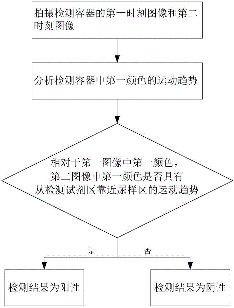
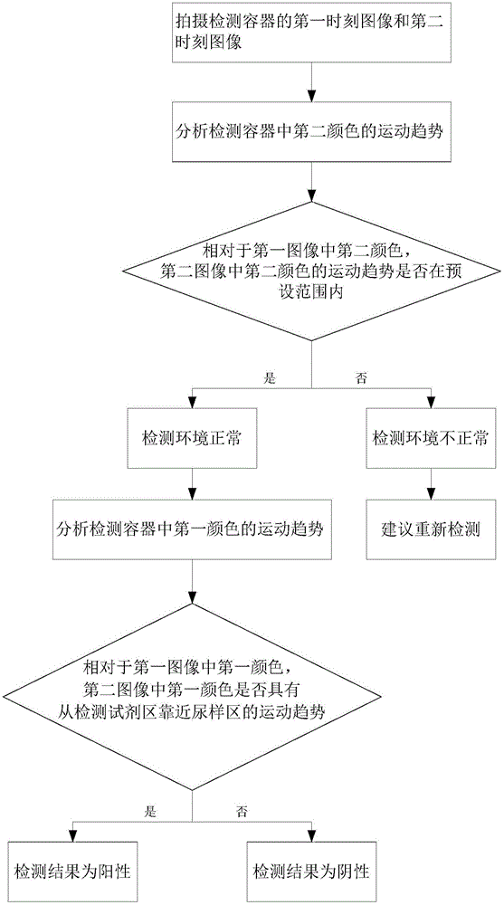
[0030] The invention proposes a cancer detection device, which includes an imaging device and a processing device.

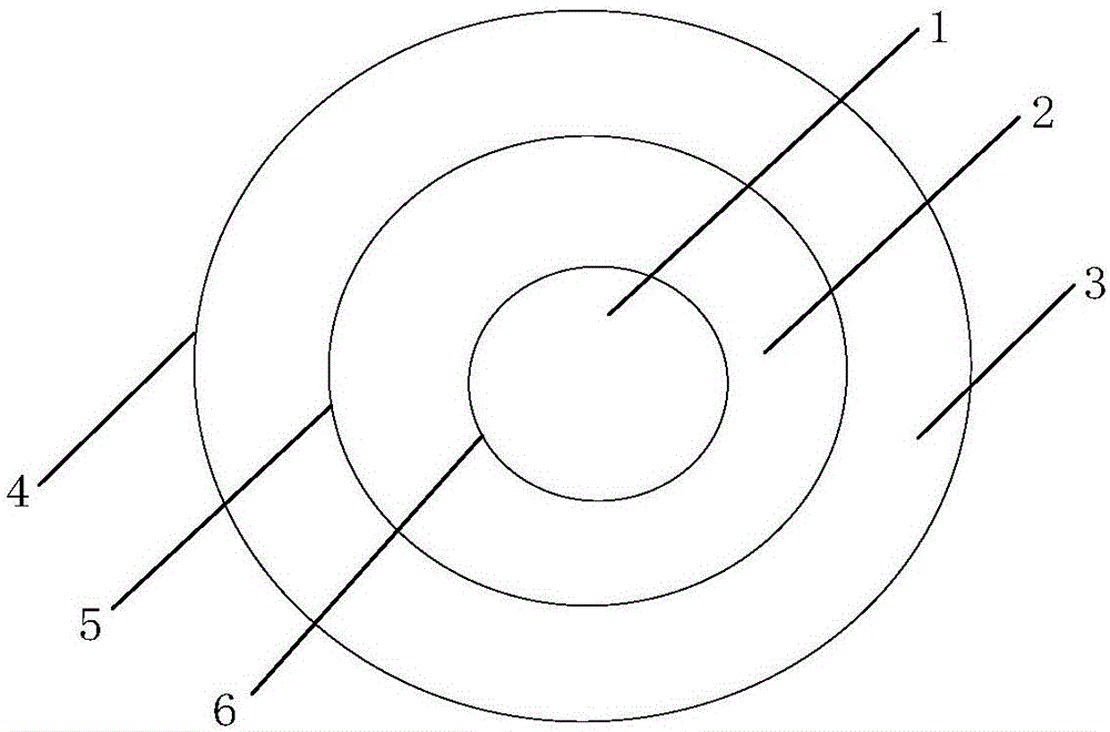
[0031] The imaging device is used to photograph the detection container at the first moment and the detection container at the second moment to obtain an image at the first moment and an image at the second moment respectively. The detection container includes a urine sample area and a detection reagent area, the urine sample area and the detection reagent ar...


PUM


Abstract

The present invention relates to cancer detection equipment, which comprises an imaging device and a treatment device, wherein the imaging device is used for shooting a detection container at a first time and the detection container at a second time to respectively obtain a first time image and a second time image, the detection container comprises a urine sample region and a detection reagent region, a detected urine sample and a detection reagent are respectively added to the urine sample region and the detection reagent region, the first nematode contained in the detection reagent is stained with a first color and has movement activity, the treatment device is used for analyzing the movement tendency of the first color, and compared with the first color in the first image, the detection result is determined as the cancer-positive result if the first color in the second image has the move tendency from the detection reagent region to the region close to the urine region, and the detection result is determined as the cancer-negative result if the first color in the second image has the movement tendency from the detection reagent region to the region away from the urine sample region. The cancer detection equipment of the present invention can be used as the low-cost, rapid and effective early cancer screening equipment.

Description

technical field [0001] The invention relates to the field of medical instruments, in particular to a cancer detection device. Background technique [0002] According to statistics, there are about 25,000 hospitals in China, of which there are less than 1,200 tertiary and first-class hospitals, and about 80% of them are concentrated in the eastern region. However, the medical resources in the central and western regions are seriously insufficient. In the vast central and western regions, which account for 80% of the country's land area and 60% of the total population, the number of doctors per thousand people is less than 1.5, which is only about 40% of that in the eastern region. This disparity in medical resources has led to the early detection and timely treatment of cancer patients in the central and western regions far behind the eastern region, which is not conducive to the early detection and treatment of cancer. [0003] Existing technologies mainly use medical imag...

Claims


Application Information

Patent Timeline
IPC IPC(8): G01N21/25
Inventor 张娟
Owner 深圳市搜罗盟环保科技有限公司
Learn More