Open quantitative analysis method and system based on medical image

A technology of medical imaging and analysis methods, applied in image analysis, image enhancement, image data processing, etc., can solve problems such as auxiliary diagnosis, prognosis and lack of prediction accuracy, and improve the effect of diagnosis

Inactive Publication Date: 2017-01-25
BEIJING DIGITAL PRECISION MEDICAL TECH CO LTD
View PDF5 Cites 30 Cited by
  • Summary
  • Abstract
  • Description
  • Claims
  • Application Information

AI Technical Summary

Problems solved by technology

[0006] When using medical images for diagnosis, in the prior art, the traditional method of visually inspecting images is mainly used, and judgments are made only by experience.

Method used


Image

  • Open quantitative analysis method and system based on medical image
  • Open quantitative analysis method and system based on medical image

Examples


specific Embodiment approach

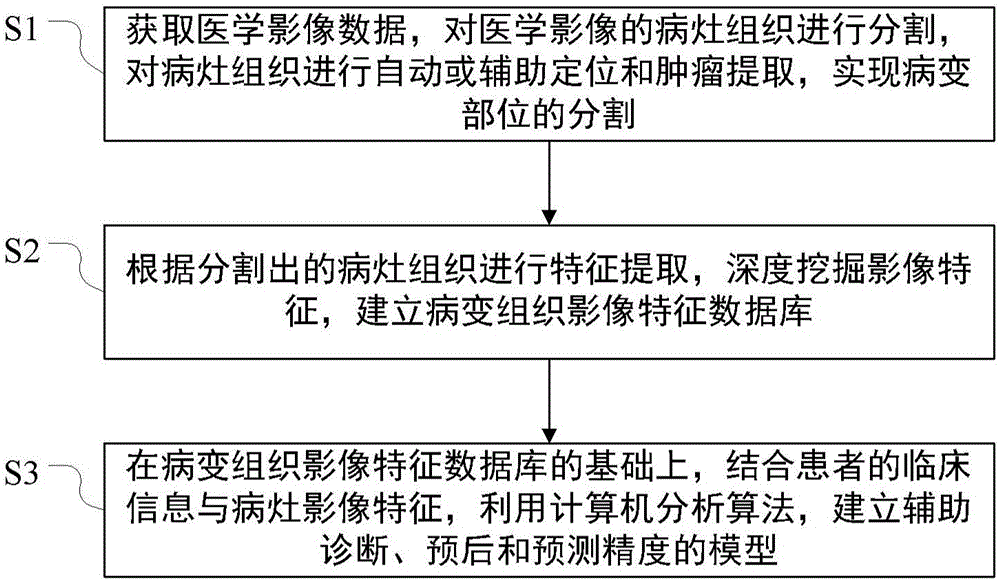
[0032] A specific embodiment of the present invention provides an open quantitative analysis method based on medical images, comprising the following steps:

[0033] S1. Obtain medical imaging data, segment the lesion tissue of the medical image, perform automatic or assisted positioning and tumor extraction on the lesion tissue, and realize the segmentation of the lesion;

[0034] Among them, an automatic segmentation algorithm based on random walk is used to segment the lesion tissue in the database. Based on the initial tumor-containing area, the algorithm adopts a recursive histogram equalization to realize the rough extraction of tumors, and then selects the foreground and background of the seed points.

[0035] It is known from experiments that the four sub-points can get the best effect, and the required tumor can be obtained through the calculation method of random walk, which can achieve good real-time and robustness of segmentation.

[0036] S2. Perform feature extr...


PUM


Abstract

The invention provides an open quantitative analysis method based on a medical image. The method comprises the steps that 1, medical image data is acquired, focus tissue in the medical images is segmented, automatic or auxiliary positioning and tumor extraction are conducted on the focus tissue, and segmentation of the diseased part is achieved; 2, feature extraction is conducted on the segmented focus tissue, image features are mined, and a focus tissue image feature database is built; 3, a model for assisting in diagnosis, prognosis and prediction precision is built on the basis of the focus tissue image feature database by combining clinical information and focus image features of a patient through a computer analysis algorithm. According to the analysis method, by applying a big data analysis method to image omics, diagnosis of a doctor is better assisted.

Description

technical field [0001] The present invention relates to the technology of radiomics-assisted diagnosis, in particular to an open quantitative analysis method and an open quantitative analysis system. Background technique [0002] In recent years, in the field of medical imaging, a large number of medical images has brought new research opportunities: [0003] a) they can give robust and reliable medical image analysis reports based on a large number of images; [0004] b) They can develop more advanced methods that rely on many samples and examples, such as data mining and machine learning, which can be introduced into the field of medical image analysis. [0005] The rapid development of medical imaging has promoted comprehensive analysis methods of radiomics medical images. Radiomics generally refers to methods for advanced characterization using computed tomography (CT), positron emission tomography (PET) or magnetic resonance imaging (MRI) high-quality fluorescence ima...

Claims


Application Information

Patent Timeline
IPC IPC(8): G06F19/00G06T7/00G06T7/11
CPCG06T7/0012G06T2207/10081G06T2207/10104G06T2207/30096
Inventor 迟崇巍王丽
Owner BEIJING DIGITAL PRECISION MEDICAL TECH CO LTD
Why Eureka
Try Eureka