Depth-learning-based eye-fundus image processing method, device and system
A fundus image and deep learning technology, which is applied in image data processing, image enhancement, image analysis, etc., can solve the problems of strong subjectivity of analysis results and high labor costs, and achieve unacceptability in solving time, reducing incompleteness, Analysis results are objective and accurate
- Summary
- Abstract
- Description
- Claims
- Application Information
AI Technical Summary
Problems solved by technology
Method used
Image
Examples
Embodiment 1
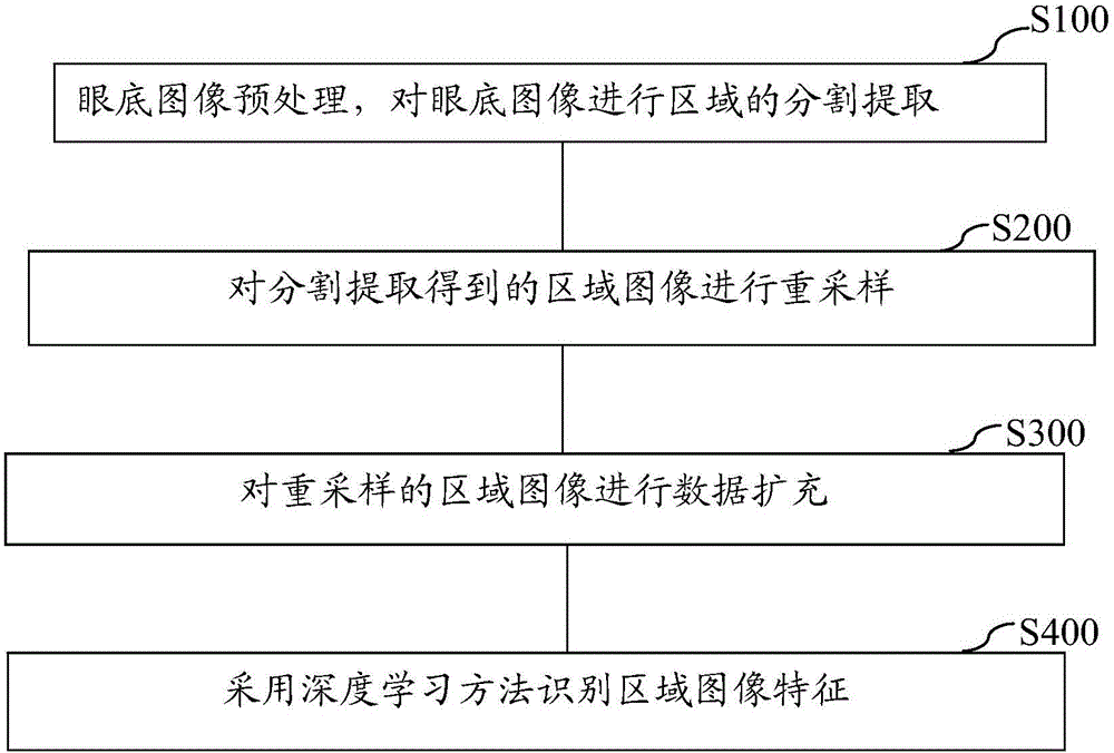
[0036] like figure 1 Shown is the fundus image processing method based on deep learning in this embodiment, which is characterized in that it includes steps S100 to S400:
[0037] S100: Firstly, the fundus image is preprocessed, and the region of the fundus image is segmented and extracted;
[0038] S200: Then resampling the region image obtained by segmentation and extraction;
[0039] S300: Perform data expansion on the resampled regional image;
[0040] S400: Using a deep learning method to identify regional image features.
Embodiment 2
[0042] On the basis of Embodiment 1, wherein step S100 includes the following steps: First, in order to eliminate the differences between images caused by different illumination conditions and camera resolutions, calculate the color average value of the entire fundus image area, and subtract any pixel of the fundus image Go to the average color. Secondly, the multi-level parabola is used to describe the shape of the fundus blood vessels, and the centerline of the fundus blood vessels is identified. Then, the position of the optic disc region (the place where the blood vessels gather) is obtained by using the direction of the blood vessels, and the optic disc region is obtained by ellipse fitting. Taking the center of the optic disc as the origin, and expanding outward by 50 pixels from the origin to the farthest edge of the optic disc as the radius, the optic cup area and the periapapillary atrophic area (PPA) were extracted.
[0043] Preferably, step S200 includes the follow...
Embodiment 3
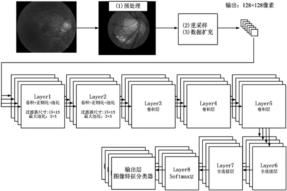
[0046] like figure 2 As shown, it is a flow chart of the training process of the convolutional neural network. First, the fundus image is preprocessed, and then resampling and data expansion are performed according to the method in Example 2. The trained convolutional neural network recognizes and recognizes the fundus image. analyze.
[0047] like figure 2 As shown, the convolutional neural network architecture includes 5 convolutional layers with weights and 2 fully connected layers, and the input layer is the image generated by resampling in the image preprocessing step. Connected to the input layer are 5 convolutional layers (Convolutional Layers). After the first and second convolutional layers are convoluted, the ReLUS (rectified linearunits) function is used for processing to speed up the training of the neural network; and then Local Response Normalization (Formula 1) is performed to prevent Overfitting, and finally Max Pooling (MaxPooling).
[0048] given Repr...
PUM
 Abstract
Description
Claims
Application Information
 - R&D
- Intellectual Property
- Life Sciences
- Materials
- Tech Scout
- Unparalleled Data Quality
- Higher Quality Content
- 60% Fewer Hallucinations
Browse by: Latest US Patents, China's latest patents, Technical Efficacy Thesaurus, Application Domain, Technology Topic, Popular Technical Reports.
© 2025 PatSnap. All rights reserved.Legal|Privacy policy|Modern Slavery Act Transparency Statement|Sitemap|About US| Contact US: help@patsnap.com


