Device and method for coronary artery calcification detection and quantification in CTA image

A CT image and quantification method technology, applied in the field of medical image data processing, can solve problems such as difficulty, inaccurate algorithm detection and quantification results, cumbersome training process, etc.

Inactive Publication Date: 2017-02-22
TIANJIN UNIV
View PDF3 Cites 55 Cited by
  • Summary
  • Abstract
  • Description
  • Claims
  • Application Information

AI Technical Summary

Problems solved by technology

[0008] In summary, these calcification detection and quantification methods generally have problems such as inaccurate algorithm detection and quantification results, high difficulty, and cumbersome training process.

Method used


Image

  • Device and method for coronary artery calcification detection and quantification in CTA image
  • Device and method for coronary artery calcification detection and quantification in CTA image
  • Device and method for coronary artery calcification detection and quantification in CTA image

Examples


Embodiment Construction

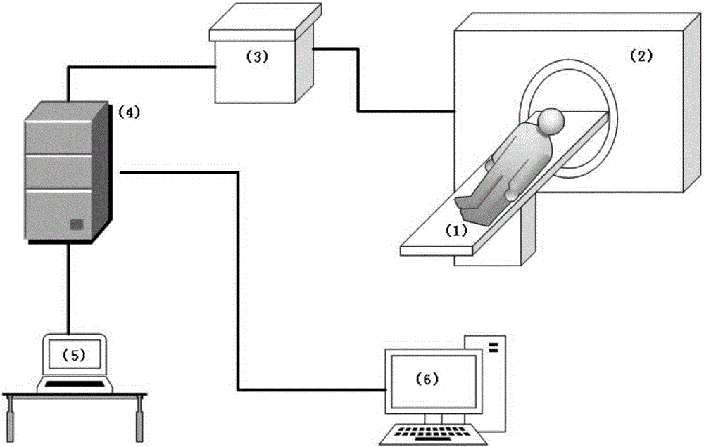
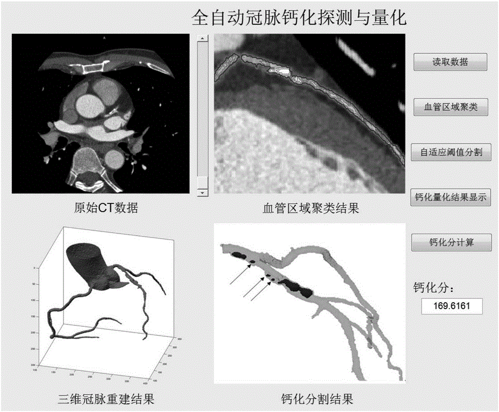
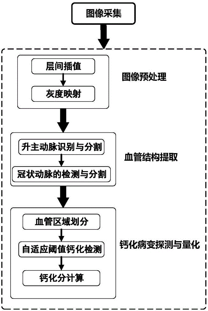
[0083] The present invention proposes a fully automatic calcified plaque detection, segmentation and quantification method based on fuzzy superpixel clustering in CTA data. Firstly, the low threshold region growth automatically selected by the seed point is used to obtain the coronary artery region containing calcified plaque; and then According to the Euclidean distance between the pixels in the region, the gray difference uses the fuzzy C-means clustering algorithm to divide the above blood vessel region into a limited number of superpixel sets; finally, a threshold selection method based on the gray histogram is used to select the superpixel set Screening is carried out to obtain the final calcified plaque detection and quantification results, and the calculation of vascular calcification scores is completed based on the segmentation results.

[0084] An automatic method for detecting and quantifying calcified plaque in CTA data, the method comprising:

[0085] Step 1. Afte...


PUM


Abstract

The present invention relates to an image processing technique for effectively suppressing noise by providing a fully automatic calcification patch detection, segmentation and quantization apparatus and method based on fuzzy super pixel clustering in CTA data. The technical scheme adopted in the present invention is the method for the enhancement of coronary artery calcification detection and quantification in CT image. The method comprises the following steps: first, using a seed point to automatically choose a low threshold region to grow and obtain a coronary artery region containing the calcification patch; then using the fuzzy C-means clustering algorithm to divide the above-mentioned blood vessel region into a finite number of super-pixel sets according to the Euclidean distances among the pixel points and the gray scale differences of the pixels in the region; and finally, using a threshold selection method based on gray histogram to screen the super pixel sets to obtain the final measurement and quantification result of the calcification patch, and completing the calcification score calculation of the blood vessel according to the segmentation result. The invention is used mainly in image processing.

Description

technical field [0001] The present invention relates to image processing technology, in particular to medical image data processing technology in enhanced computed tomography (Computed Tomography, CT) images, especially a method for detecting and quantifying coronary artery calcified plaques in enhanced CT image data , which is a method for the detection and quantification of coronary artery calcification in enhanced CT images. Background technique [0002] Coronary heart disease refers to the stenosis or blockage of the vascular lumen due to atherosclerosis, inflammation, embolism and other lesions in the coronary arteries, which in turn causes myocardial ischemia, hypoxia or necrosis, especially when the atherosclerotic plaque ruptures suddenly It may cause sudden blockage of the cardiovascular system and sudden death of the patient. It is one of the diseases with the highest fatality rate in the world. Medical research shows that the detection and quantification of coron...

Claims


Application Information

Patent Timeline
Patent Type & Authority Applications(China)
IPC IPC(8): G06T7/00G06T7/11G06T7/136
Inventor 陈晓冬赵聪王丽瑶张佳琛汪毅郁道银
Owner TIANJIN UNIV
Why Eureka
Try Eureka