Reconstruction and numerical value calibration method for medical dual-energy CT electron density image

A technology of electron density and calibration method, applied in the fields of radiological diagnostic image/data processing, medical science, computer tomography scanner, etc., can solve the problems of inaccurate numerical value, cumbersome process, high acquisition cost, and achieve improved accuracy and easy engineering realization. Effect

Active Publication Date: 2017-03-08
SHANDONG UNIV
View PDF6 Cites 10 Cited by
  • Summary
  • Abstract
  • Description
  • Claims
  • Application Information

AI Technical Summary

Problems solved by technology

[0004] In the prior art, the acquisition cost of phantoms for human tissue electron density images is high, the process is cumbersome, and the values ​​are inaccurate, and the mainstream large-scale digital medical equipment manufacturers at home and abroad lack practical and accurate electron density CT / DECT image reconstruction and numerical calibration methods, so far do not directly provide electron density images in their DECT products

Method used


Image

  • Reconstruction and numerical value calibration method for medical dual-energy CT electron density image
  • Reconstruction and numerical value calibration method for medical dual-energy CT electron density image
  • Reconstruction and numerical value calibration method for medical dual-energy CT electron density image

Examples


Embodiment 1

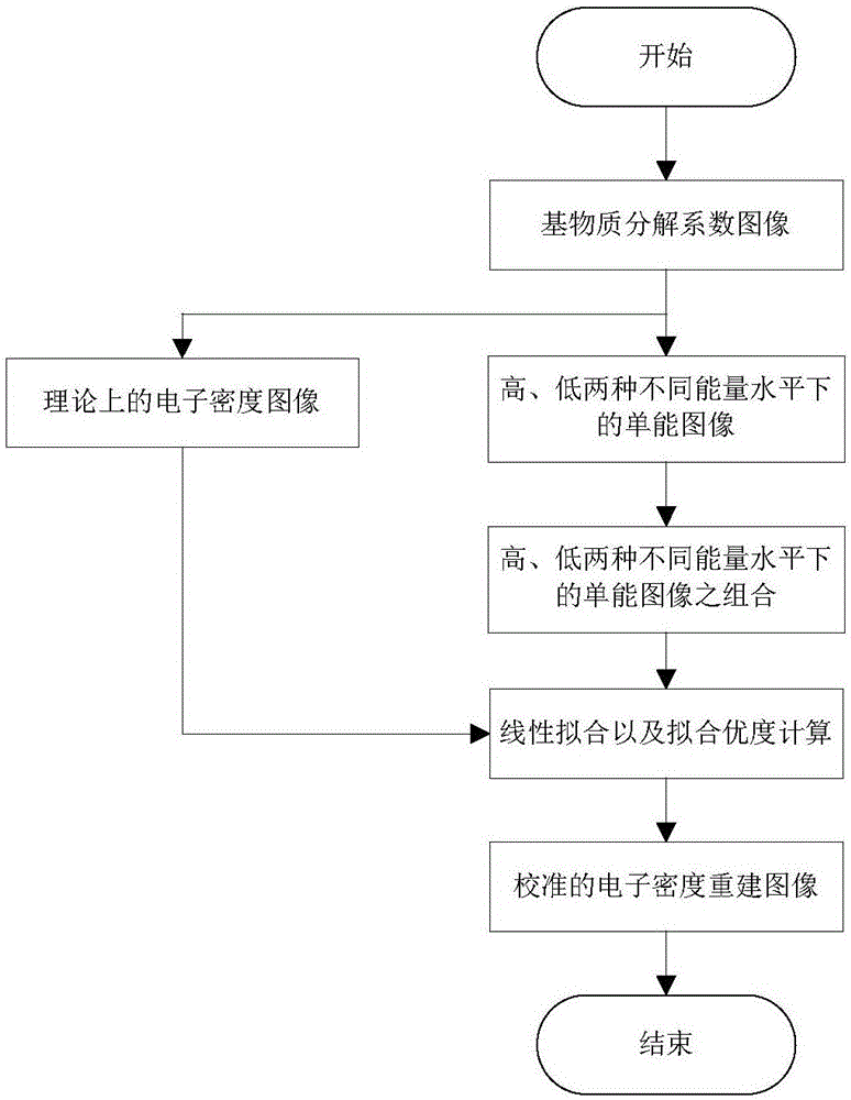
[0033] like figure 1 shown.

[0034] A method for reconstruction and numerical calibration of medical dual-energy CT electron density images, comprising the following steps:

[0035] S1) Calculate matrix decomposition coefficient image b 1 ,...,b n ;

[0036] The DECT equipment obtains the matrix material density image, and the matrix material density image is denoted as I 1 ,...,I n ; According to the following formula to calculate the matrix material decomposition coefficient image b 1 ,...,b n ; Among them, ρ i Indicates the material density of the i-th base material; the unit of material density is g / cm 3 ;n=3;

[0037] S2) Calculate the theoretical electron density image ρ e and monoenergetic images at two different energy levels, high and low;

[0038] Theoretical electron density image Among them, ρ ei = 2·ρ i·Z i / A i is the electron density of the i-th base substance, Z i and A i respectively represent the atomic number and atomic weight of the i-...

Embodiment 2

[0050] The reconstruction and numerical calibration method of the medical dual-energy CT electron density image as described in Example 1, the difference is that the matrix material density image is obtained by a DECT imaging workstation.


PUM


Abstract

The invention relates to a reconstruction and numerical value calibration method for a medical dual-energy computed-tomography (CT) electron density image. Reconstruction and numerical value calibration are carried out on a medical dual-energy-CT (DECT) electron density image according to a basal principle of base-material-decomposition DECT imaging as well as a linear relationship between combination of X-ray linear attenuation coefficients at a high energy level and a low energy level and a theoretical electron density value. When the method provided by the invention is applied to the tumor radiation therapy field for clinical application, the precision of target region positioning and accuracy of exposure dose calculation of a radiation therapy planning system can be improved.

Description

technical field [0001] The invention relates to a reconstruction and numerical calibration method of a medical dual-energy CT electron density image, belonging to the technical field of medical dual-energy CT. Background technique [0002] Radiotherapy (Radiation Therapy) is a local treatment method that uses radiation to treat tumors, and is one of the main means of treating malignant tumors. Among them, the electron density image reconstruction and numerical calibration of the human tissue in the lesion are directly related to the accuracy of the target positioning and the accuracy of the radiation dose calculation of the Radiation Therapy Planning System. Therefore, modern tumor radiotherapy needs to measure the electron density and distribution of human tissue in the lesion. At present, CT (Computed Tomography; computerized tomography) scan phantom is widely used clinically to obtain the electron density and distribution of human tissue in lesion. That is, make a phant...

Claims


Application Information

Patent Timeline
Patent Type & Authority Applications(China)
IPC IPC(8): A61B6/00A61B6/03
CPCA61B6/03A61B6/52
Inventor 孙丰荣秦娇娇张明强刘炜仲海刘芳蕾金培培于海涛王丽新
Owner SHANDONG UNIV
Why Eureka
Try Eureka