
Lung detection method and device based on PET/CT image features

A CT image and detection method technology, applied in the field of medical digital image processing, can solve problems such as incompleteness, and achieve the effect of improving accuracy and sensitivity

Inactive Publication Date: 2017-03-22
CAPITAL UNIVERSITY OF MEDICAL SCIENCES
View PDF2 Cites 23 Cited by
  • Summary
  • Abstract
  • Description
  • Claims
  • Application Information

AI Technical Summary

Problems solved by technology

However, at present, there is no perfect solution for judging tumor properties and lesion status based on PET / CT image features.

Method used


Image

  • Lung detection method and device based on PET/CT image features
  • Lung detection method and device based on PET/CT image features
  • Lung detection method and device based on PET/CT image features

Examples


Embodiment Construction

[0053] In order to make the purpose, technical solutions and advantages of the embodiments of the present invention clearer, the technical solutions in the embodiments of the present invention will be clearly and completely described below in conjunction with the drawings in the embodiments of the present invention. Obviously, the described embodiments are only It is a part of embodiments of the present invention, but not all embodiments. The components of the embodiments of the invention generally described and illustrated in the figures herein may be arranged and designed in a variety of different configurations. Accordingly, the following detailed description of the embodiments of the invention provided in the accompanying drawings is not intended to limit the scope of the claimed invention, but merely represents selected embodiments of the invention. Based on the embodiments of the present invention, all other embodiments obtained by those skilled in the art without making...


PUM


Abstract

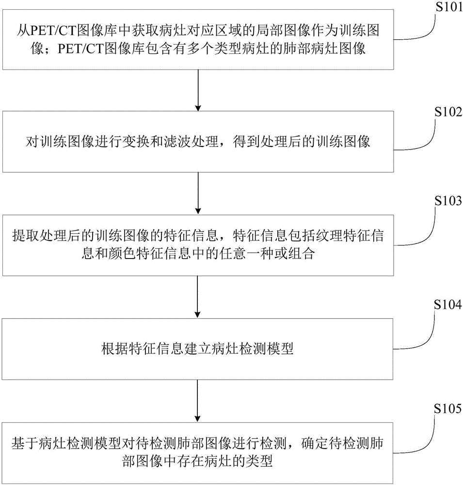
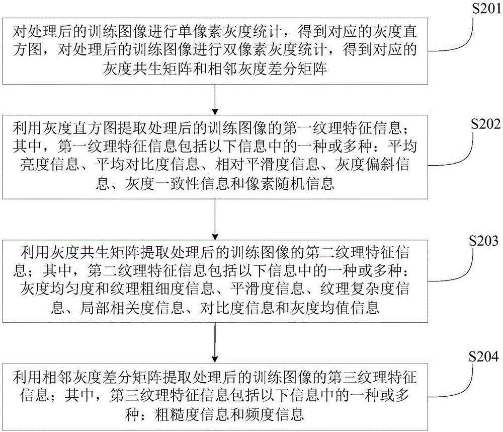
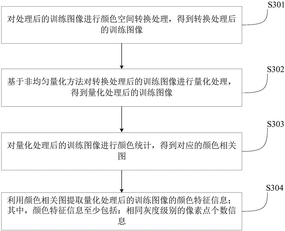
The invention provides a lung detection method and device based on PET / CT image features. The method comprises the steps that the local image of a region corresponding to a lesion is acquired from a PET / CT image library as a training image; the PET / CT image library comprises the lung lesion images of multiple types of lesions; the training image is transformed and filtered to acquire a processed training image; the feature information of the processed training image is extracted, wherein the feature information comprises one or combination of texture feature information and color feature information; a lesion detection model is established according to the feature information; and a lung image to be detected is detected based on the lesion detection model to determine the type of the lesion in the lung image to be detected. An image processing technology is used to carry out lesion analysis and identification on a PET / CT image, which improves the accuracy and sensitivity of lesion identification.

Description

technical field [0001] The present invention relates to the technical field of medical digital image processing, in particular to a method and device for lung detection based on PET / CT image features. Background technique [0002] Lung cancer is one of the deadliest cancers worldwide. PET / CT (positron emission tomography / computer tomography, positron emission tomography / computer tomography), as a non-innovative imaging diagnostic technology, has made great progress in the application of lung cancer diagnosis and preoperative staging in recent years . Compared with traditional CT (computertomography, computerized tomography), MRI (Magnetic Resonance Imaging, magnetic resonance imaging) or pure PET (positron emission tomography, positron emission tomography), the advantages of PET / CT technology are very obvious. [0003] In terms of detecting and locating tumor tissue using PET / CT images, there have been some techniques that apply threshold judgment and SUV value comparison...

Claims


Application Information

Patent Timeline
Patent Type & Authority Applications(China)
IPC IPC(8): G06T7/00G06T7/41G06T7/90
CPCG06T7/0012G06T2207/10024G06T2207/10081G06T2207/10104G06T2207/20081G06T2207/30061G06T2207/30096
Inventor 郭秀花马圆陈斯鹏田思佳
Owner CAPITAL UNIVERSITY OF MEDICAL SCIENCES
Learn More