Method and apparatus for lung segmentation in medical image

A medical image and image technology, applied in image analysis, image enhancement, image data processing, etc., can solve problems such as being easily affected by rib boundaries, achieve high work efficiency, improve accuracy, and reduce deviations

Active Publication Date: 2017-05-03
SHANGHAI UNITED IMAGING HEALTHCARE
View PDF3 Cites 14 Cited by
  • Summary
  • Abstract
  • Description
  • Claims
  • Application Information

AI Technical Summary

Problems solved by technology

But this method is to find the boundary on two-dimensional information, which is easily affected by the rib boundary, resulting in a jagged segmentation result

Method used


Image

  • Method and apparatus for lung segmentation in medical image
  • Method and apparatus for lung segmentation in medical image
  • Method and apparatus for lung segmentation in medical image

Examples


Embodiment Construction

[0051] The present invention will be further described below in conjunction with the accompanying drawings and embodiments.

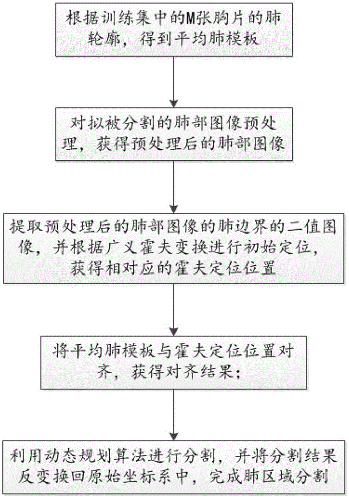
[0052] see figure 1 -3, a method for lung segmentation in a medical image (such as a DR image or an X-ray image) according to an embodiment of the present invention, comprising the following steps:

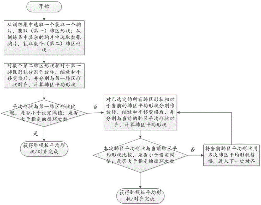
[0053] According to the lung contours of the M chest films in the training set, an average lung template is obtained, where M is an integer greater than or equal to 2;

[0054] Obtain the lung image to be segmented;

[0055] Preprocessing the lung image to be segmented to obtain the preprocessed lung image;

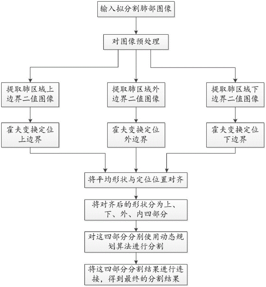
[0056] Extract the binary image of the lung boundary of the preprocessed lung image, and perform initial positioning according to the generalized Hough transform to obtain the corresponding Hough positioning position;

[0057] Align the average lung template with the Hough positioning position to obtain the Hough positioning result;

[0058] The dy...


PUM


Abstract

The invention discloses a method and an apparatus for lung segmentation in a medical image. The method comprises the following steps of obtaining an average lung template according to lung contours of M chest films in a training set, wherein M is an integer greater than or equal to 2; preprocessing a lung image to be segmented to obtain a preprocessed lung image; extracting a binary image of a lung boundary of the preprocessed lung image, and performing initial locating according to generalized Hough transform to obtain a corresponding Hough locating position; aligning the average lung template to the Hough locating position to obtain an alignment result; performing segmentation by utilizing a dynamic planning algorithm, and inversely transforming a segmentation result to be in an original coordinate system, thereby finishing lung region segmentation. According to the method and the apparatus, the lung region to be segmented is located through the Hough transform, and the locating position is aligned through an average lung region shape to obtain an initial contour of the lung, so that a deviation of the initial position and the actual position of the lung is reduced to the maximum extent and the accuracy of segmentation is improved.

Description

[0001] 【Technical field】 [0002] The invention relates to the field of medical image segmentation, in particular to a method and device for lung segmentation in medical images. [0003] 【Background technique】 [0004] Lung segmentation is an important link in digital chest radiograph image processing. The quality of the segmentation result directly affects the subsequent detection and analysis of the lesion. The biggest drawback of chest radiographs is tissue overlap. The higher the density of the tissues in the human body structure, the stronger the ability to absorb X-rays. Therefore, the ribs in the chest absorb more X-rays and appear as white shadows in the image; because the lungs contain air, they are less dense and absorb X-rays. Less, so dark shadows appear on the image. Because of this, jagged segmentation results are prone to appear when segmenting lung boundaries. Moreover, by observing the image, it can be found that there are corner points at the lower boundar...

Claims


Application Information

Patent Timeline
Patent Type & Authority Applications(China)
IPC IPC(8): G06T7/10G06T7/136
CPCG06T2207/20081G06T2207/30061
Inventor 丛琳郭薇李强
Owner SHANGHAI UNITED IMAGING HEALTHCARE
Why Eureka
Try Eureka