A multi-frequency electromagnetic tomography method for the detection of intracerebral hemorrhage

A technology of electromagnetic tomography and detection coils, which is applied in the fields of application, diagnostic recording/measurement, medical science, etc., and can solve problems such as unfavorable long-term continuous monitoring, high price, and containing radioactive sources

Active Publication Date: 2020-10-20
TIANJIN UNIV
View PDF5 Cites 0 Cited by
  • Summary
  • Abstract
  • Description
  • Claims
  • Application Information

AI Technical Summary

Problems solved by technology

However, these imaging methods are expensive and contain radioactive sources, which are not conducive to long-term continuous monitoring

Method used


Image

  • A multi-frequency electromagnetic tomography method for the detection of intracerebral hemorrhage
  • A multi-frequency electromagnetic tomography method for the detection of intracerebral hemorrhage
  • A multi-frequency electromagnetic tomography method for the detection of intracerebral hemorrhage

Examples


Embodiment Construction

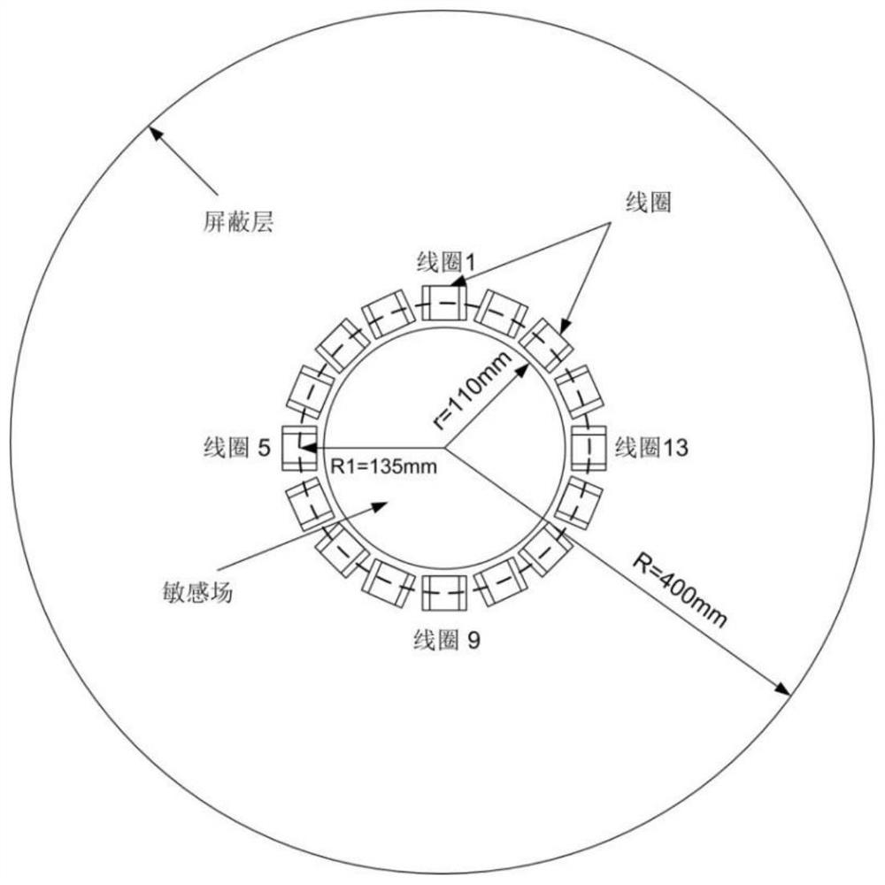
[0018] The multi-frequency imaging method of electromagnetic tomography is based on the characteristics that the dielectric characteristic parameters of biological tissues change with frequency, and according to the characteristics that the phase shift of the detection voltage changes linearly with frequency and conductivity, the imaging results of a single tissue of cerebral hemorrhage can be reconstructed, both It can overcome the shortcoming that the time difference method cannot obtain the object field information before the occurrence of cerebral hemorrhage, and can eliminate the artifacts in the imaging results of cerebral hemorrhage obtained by the dual-frequency frequency difference method. The multi-frequency imaging method obtains the detection voltage phase shift generated by the brain tissue containing cerebral hemorrhage on the detection coil at different frequencies, separates the detection voltage phase shift generated by a single tissue on the detection coil, and...


PUM


Abstract

The invention discloses a multi-frequency electromagnetic tomographic method for detection of cerebral hemorrhage. Multiple coils are distributed on the periphery of a detected area, exciting coils are sequentially powered up with alternating excitation currents at different frequencies, detection coils around a detected area produce induced voltages sequentially at different frequencies, and the target cerebral hemorrhage image is reconstructed in steps as follows: the detection voltage difference produced by a brain suffering from cerebral hemorrhage on the detection coils is calculated; phase shift, caused by the brain suffering from cerebral hemorrhage when the frequency of excitation currents is fi, of detection voltage on the detection is calculated; the formula is solved with a Tikhonov regularization method, the phase shift, caused by single tissue j when the reference frequency of the excitation currents is f1, of the detection voltage on the detection coils is acquired; the phase difference, caused by cerebral hemorrhage tissue when the frequency of the excitation currents is between fn and f1, of the detection voltage on the detection coils is calculated; the electric conductivity distribution of the cerebral hemorrhage tissue is solved.

Description

technical field [0001] The invention belongs to the technical field of biological imaging and relates to a multi-frequency electromagnetic tomography imaging method. [0002] technical background [0003] At present, the medical imaging methods used clinically to detect cerebral hemorrhage include CT, MRI and so on. However, these imaging methods are expensive and contain radioactive sources, which are not conducive to long-term continuous monitoring. Electromagnetic tomography is an electrical tomography technology based on the principle of electromagnetic induction. Because the electromagnetic field can penetrate the skull with low conductivity, and its non-contact, radiation-free, and low-cost characteristics, it can realize the diagnosis of cerebral hemorrhage and other lesions. Long-term continuous monitoring has great development prospects. [0004] When electromagnetic tomography technology is used in the detection of cerebral hemorrhage, there are two methods: time ...

Claims


Application Information

Patent Timeline
Patent Type & Authority Patents(China)
IPC IPC(8): A61B5/05A61B5/00
CPCA61B5/05A61B5/7203
Inventor 谭超肖志利董峰
Owner TIANJIN UNIV
Why Eureka
Try Eureka