Low-dose X-ray CT image reconstruction method

A CT image and X-ray technology, applied in the field of low-dose X-ray CT image reconstruction, can solve the problems of reduced CT image resolution, inability to meet clinical CT real-time imaging, and loss of original image details.

Active Publication Date: 2017-05-31
XI AN JIAOTONG UNIV +1
View PDF3 Cites 11 Cited by
  • Summary
  • Abstract
  • Description
  • Claims
  • Application Information

AI Technical Summary

Problems solved by technology

When reconstructing CT images with the same pixel size, the time required for iterative reconstruction methods based on statistical models is often much longer than that of classical analytical reconstruction methods, so it still cannot meet the needs of clinical CT real-time imaging
[0006] However, the traditional analytical reconstruction method based on projection data filtering does not make full use of the inherent statistical information of the data, and the original detail information of the image is often lost in the process of projection data restoration, thereby reducing the resolution of the corresponding CT image.

Method used


Image

  • Low-dose X-ray CT image reconstruction method
  • Low-dose X-ray CT image reconstruction method
  • Low-dose X-ray CT image reconstruction method

Examples


Embodiment 1

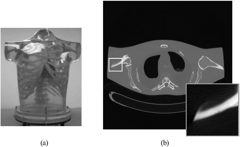
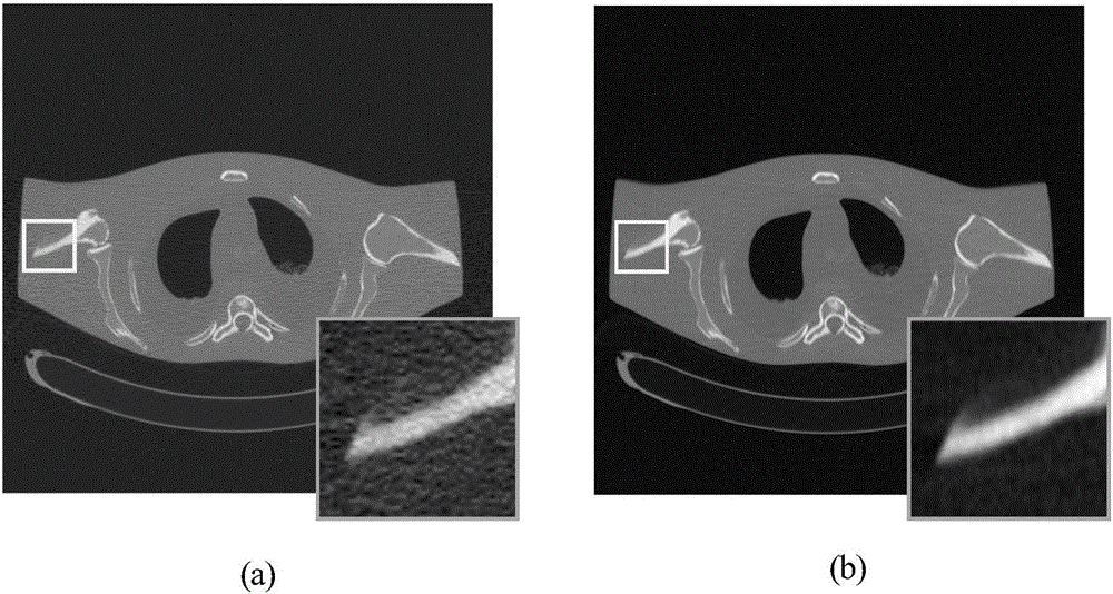
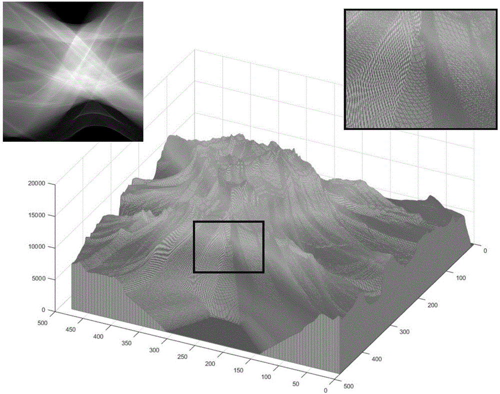
[0092] use as figure 2 The shown phantom image is used as the computer simulation experiment object of the present invention. The size of the phantom image is set to 512×512, the distances from the X-ray source of the simulated CT equipment to the rotation center and the detector are 1361.2mm and 615.18mm respectively, and the sampling value of the rotation angle is 1160 between [0,2π]. The angle corresponds to 672 detector units, and the size of the detector unit is 1.85mm. The fragment of the chord diagram data is as follows Figure 4 shown. Choose slice-plane approximation features for chordal graphs (such as Figure 5 Shown) is a priori, and the present invention comprises the following steps successively:

[0093] Step S1: Obtain the imaging system parameters of the CT equipment and the projection data p under the low-dose CT scanning protocol; the imaging system parameters of the acquired CT equipment include X-ray incident photon intensity I 0 , the variance σ of t...

Embodiment 2

[0149] use as figure 2 The shown phantom image (chord figure) is used as the computer simulation experiment object of the present invention. The size of the phantom image is set to 512×512, the distances from the X-ray source of the simulated CT equipment to the rotation center and the detector are 1361.2mm and 615.18mm respectively, and the sampling value of the rotation angle is 1160 between [0,2π]. The angle corresponds to 672 detector units, and the size of the detector unit is 1.85mm. The fragment of the chord diagram data is as follows Figure 4 shown. Select the smoothing prior of the CT image (such as Image 6 Shown) is a priori, and the present invention comprises the following steps successively:

[0150] Step S1: Obtain the imaging system parameters of the CT equipment and the projection data p under the low-dose CT scanning protocol; the imaging system parameters of the acquired CT equipment include X-ray incident photon intensity I 0 , the variance σ of the e...


PUM


Abstract

The present invention provides a low-dose X-ray CT image reconstruction method. The method comprises: obtaining imaging system parameters of a CT device and projection data in the low-dose CT scanning protocol; constructing a statistics generation model of the chordal graph data through the statistical law of the imaging process; constructing the priori statistic model of the chordal graph data; constructing a complete statistic model, and converting a model to a chordal graph data recovery model according to the maximum posterior estimation method; applying the chordal graph data recovery model, and obtaining estimated chordal graph data and other statistical variables; and performing CT image reconstruction according to the obtained chordal graph data to obtain and output a CT graph according to the obtained chordal graph data. The objective is to establish a high-quality chordal graph data recovery model and method based on the chordal graph data generation principle and the statistical prior therein so as to realize the CT image high-quality reconstruction capable of greatly reducing image noise/artifact and recovering image details.

Description

technical field [0001] The invention relates to an image processing technology of medical images, in particular to a low-dose X-ray CT image reconstruction method based on statistical modeling and maximum a posteriori estimation framework. Background technique [0002] X-ray CT scanning has been widely used in clinical medical imaging diagnosis, but the excessive X-ray radiation dose in the process of CT scanning has potential risks to the human body, which may easily cause radiation damage and induce malignant tumors. On the premise of ensuring the image quality, minimizing the dose of X-rays has become one of the key technologies in the field of medical CT imaging. [0003] In order to reduce the dose of X-ray radiation, various hardware and software technologies can be used to reduce the dose of X-ray used in CT scanning. Common techniques include reducing tube current, reducing X-ray exposure time, and reducing the amount of projection data acquired (ie, sparse-angle CT...

Claims


Application Information

Patent Timeline
Patent Type & Authority Applications(China)
IPC IPC(8): G06T11/00G06T5/00
CPCG06T5/001G06T11/008G06T2211/421G06T2207/10081
Inventor 谢琦孟德宇马建华赵谦徐宗本
Owner XI AN JIAOTONG UNIV
Why Eureka
Try Eureka