Conductivity reconstruction method for biological-induction type magnetic-acoustic endoscopic imaging

A biosensing and conductivity technology, applied in the field of medical imaging, can solve problems such as difficult to apply EMAT-MI imaging

Inactive Publication Date: 2017-06-20
NORTH CHINA ELECTRIC POWER UNIV (BAODING)
View PDF2 Cites 1 Cited by
  • Summary
  • Abstract
  • Description
  • Claims
  • Application Information

AI Technical Summary

Problems solved by technology

[0004] The purpose of the present invention is to provide a conductivity reconstruction method for EMAT-MI imaging to improve the quality of reconstructed images for the disadvantages that the existing MAT-MI image reconstruction algorithm is difficult to apply to EMAT-MI imaging

Method used


Image

  • Conductivity reconstruction method for biological-induction type magnetic-acoustic endoscopic imaging
  • Conductivity reconstruction method for biological-induction type magnetic-acoustic endoscopic imaging
  • Conductivity reconstruction method for biological-induction type magnetic-acoustic endoscopic imaging

Examples


Embodiment Construction

[0038] The processing steps of the present invention include:

[0039] 1. Establish a multi-layer cavity tissue cross-sectional model:

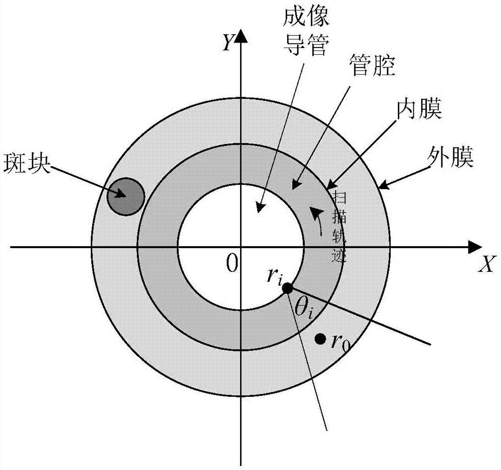
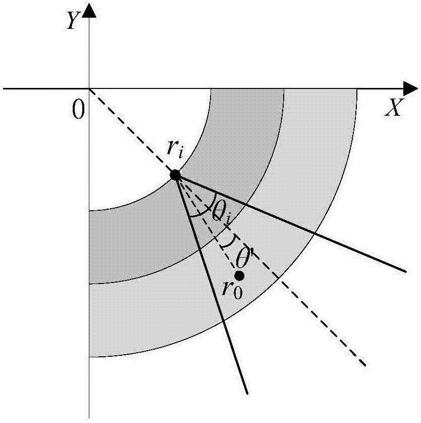
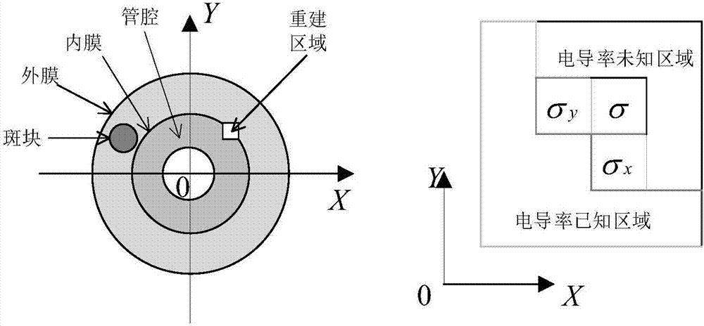
[0040] Take the blood vessel cross-section model as an example, as shown in the attached figure 1 As shown, the model includes the adventitia, intima, lumen, atherosclerotic plaque, and imaging catheter of the vessel wall. The ultrasound probe is located at the top of the imaging catheter and is used to receive the magnetoacoustic signals generated by the wall tissue. The catheter is driven by a micro motor to rotate, and the cavity tissue is scanned in a circumferential direction, and the scanning plane is perpendicular to the imaging catheter. In order to simplify the problem, the ultrasonic detector is approximated as an ideal point detector, its aperture size is ignored, and an X-Y plane Cartesian coordinate system is established on the imaging plane, with the center of the imaging catheter as the coordinate origin, and the horizontal d...


PUM


Abstract

The invention relates to a conductivity reconstruction method for bioreactor magnetic endoscopic imaging. The conductivity reconstruction method is characterized in that pretreatment including superposed averaging, filter denoising and deconvolution is carried out on original magnetic acoustic signals collected by an ultrasonic detector based on establishment of multi-layer cavity tissue cross-section model; sound source distribution at the cross section of the multi-layer cavity wall tissue is reconstructed by using the pre-processed magnetic acoustic signals; and then according to the obtained sound source distribution, a conductivity distribution graph of the tissue cross section is reconstructed. According to the conductivity reconstruction method, the conductivity distribution graph of the tissue cross section can be constructed accurately and rapidly by using the magnetic acoustic signals generated by biological tissues and thus magnetic-acoustic endoscopic imaging of the biological cavity tissues can be realized, so that accurate and reliable information can be provided for the early-stage disease diagnosis.

Description

technical field [0001] The invention relates to a method for reconstructing a conductivity distribution map of a cavity tissue cross section by using magnetoacoustic signals generated by biological tissues, and belongs to the technical field of medical imaging. Background technique [0002] Bioinductive magnetic acoustic endoscopic imaging (Endoscopic Magnetoacoustic Tomography with Magnetic Induction, EMAT-MI) is to measure and collect the electrical characteristic information of the cavity tissue from the cavity (such as the digestive tract, intestinal tract and blood vessels, etc.), and its imaging principle is The tissue to be tested is placed in a static magnetic field whose magnetic field direction is along the axis of the lumen, and at the same time, a pulsed magnetic field with the same direction as the static magnetic field and whose frequency is in the ultrasonic band is applied. Under the action of the pulsed magnetic field, an induced current will be generated in...

Claims


Application Information

Patent Timeline
IPC IPC(8): G06T5/00G06T5/50G06T7/00
CPCG06T5/50G06T7/0012G06T2207/20216G06T2207/30021G06T2207/10068G06T5/70
Inventor 孙正陈鹏
Owner NORTH CHINA ELECTRIC POWER UNIV (BAODING)
Why Eureka
Try Eureka