
A tissue graft direct suspending and fixing device

A tissue graft and fixation device technology, applied in the field of medical devices, can solve problems such as joint instability, unfavorable graft and bone tunnel healing, ligament loosening, etc., to achieve firm and reliable connection, promote quick healing, and avoid the effect of wiper effect.

Pending Publication Date: 2017-09-15
上海利格泰生物科技股份有限公司
View PDF5 Cites 2 Cited by
  • Summary
  • Abstract
  • Description
  • Claims
  • Application Information

AI Technical Summary

Problems solved by technology

If the coil length of the suspension fixation device is fixed, there will be a cavity with a length of about 6mm in the femoral tunnel, and the graft will have a pendulum effect and a bungee effect, which may cause a series of problems, such as enlarged bone tunnel, hindering the bone tunnel and grafting material healing, ligament loosening, joint instability, etc.
In short, the existing suspension fixation method of the femoral end has many disadvantages, such as the graft does not fully contact the bone tunnel, which is not conducive to the healing of the graft and the bone tunnel, and affects the treatment effect; there will be a pendulum effect and a bungee effect, resulting in a series of question

Method used


Image

  • A tissue graft direct suspending and fixing device
  • A tissue graft direct suspending and fixing device
  • A tissue graft direct suspending and fixing device

Examples


Embodiment approach 1

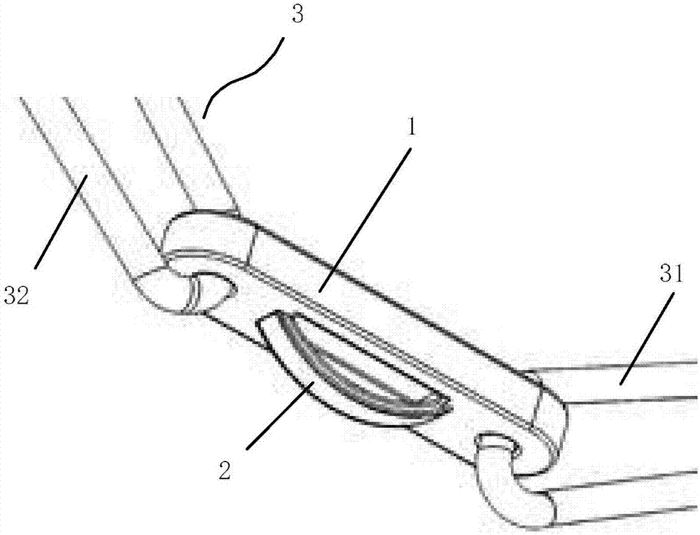
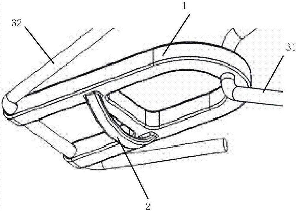
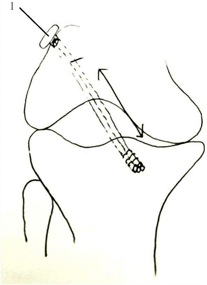
[0060] Such as image 3 Shown, the knee joint has tissue grafts that can be implanted in cruciate ligament (ACL) reconstruction and repair surgery, eg patellar tendon graft, hamstring tendon graft. Take a graft, measure the length, fold it into 4 strands, and place it on the pretensioner. After a certain tensile force, pass the transplanted tendon through the suspension rod 2, and suture the two ends of the 4 strands of tendon with medical sutures. The length is about 8cm, and the diameter is 8-9mm.

[0061] A tibial tunnel is then drilled into the tibia at a predetermined angle and distance to place the tibial graft; a femoral tunnel is also drilled at a predetermined angle and distance to place the femoral graft. The femoral tunnel was made the same diameter to accommodate thicker tendon grafts.

[0062] Connect the traction wire 31 and the turning wire 32 to the threader and pull it out from bottom to top through the outer opening of the tibial tunnel-the inner opening of...

Embodiment approach 2

[0064] The patient is positioned supine and anesthetized. First, a longitudinal incision about 2 cm long is made downward from the place 4 cm below the tibial plateau and 2 cm inside the tibial tuberosity of the affected knee to expose the insertion point of the semitendinosus and gracilis of the foot of the goosefoot, and cut off the periosteum together. After part of the semitendinosus and gracilis muscle insertions were freed, the tendon was cut off with a tendon stripper, trimmed into a 14-18cm long tendon bundle, and the muscles and excess fascia attached to the tendon were removed, and the transplanted tendon was put into the suspension. Inside the suspender 2, braid with tendon sutures and fold into 4 strands, pre-tension and stretch for 5 minutes, and set aside.

[0065] Create a bone tunnel. The tibial insertion point of the reconstructed tendon is at the midpoint of the line between the anterior medial bundle and the posterolateral bundle at the insertion point of t...


PUM


Abstract

The invention provides a tissue graft direct suspending and fixing device comprising a fixing member, a suspension rod and sewing threads. The fixing member is provided with thread penetration holes; the sewing threads are arranged in the thread penetration holes in a penetrating manner and connected with the fixing member; the suspension rod is arranged on the lower side of the fixing member. The device enables a graft to be placed in a femoral tunnel completely, promotes full contact between the graft and the femoral tunnel and promotes rapid healing.

Description

technical field [0001] The invention relates to a medical device, in particular to a tissue graft direct suspension and fixation device. Background technique [0002] The most clinically used cruciate ligament femoral end suspension fixation device is a coil metal plate device, which generally consists of a length-adjustable or non-adjustable coil, a metal attachment plate and lead wires. The principle of suspension fixation of these products is similar: the graft passes through the coil, and the metal attachment plate is suspended outside the femoral tunnel opening to achieve the purpose of femoral end fixation. The problem is that the coil has a certain length, and there must be a section of the femoral tunnel filled with coils rather than grafts. Generally, the proximal part of the femoral tunnel has a diameter smaller than that of the distal tunnel. The reason is that the proximal part of the tunnel with a smaller diameter is used to receive the coil of the fixation dev...

Claims


Application Information

Patent Timeline
IPC IPC(8): A61F2/08
CPCA61F2/0805A61F2/0811A61F2002/0817A61F2220/0008A61F2220/0075
Inventor 霍红亚
Owner 上海利格泰生物科技股份有限公司
Learn More