Lesion detection method and apparatus of eye fundus image

A fundus image and detection method technology, applied in image enhancement, image analysis, image data processing, etc., can solve the problems of difficult filling operation, low precision, and small universality, so as to improve detection accuracy, speed up detection, The effect of improving accuracy

Active Publication Date: 2017-09-19
NANJING ZHONGXING XIN SOFTWARE CO LTD
View PDF4 Cites 32 Cited by
  • Summary
  • Abstract
  • Description
  • Claims
  • Application Information

AI Technical Summary

Problems solved by technology

However, the above methods may have boundary fractures, making filling operations difficult, and thus missing some lesion areas
[0005] In general, the existing fundus image processing methods mainly have the following defects: low precision, unable to conduct accurate quantitative analysis; need fixed parameter combination, low universality, cannot perform accurate analysis for various images; require a large number of The data is used as a training sample; some algorithms are too expensive to calculate, and the calculation cost is too high, which exceeds the computing power of general computers
[0006] It can be seen that although there are a large number of research results on fundus image analysis, the processing speed or lesion area positioning effect needs to be improved.

Method used


Image

  • Lesion detection method and apparatus of eye fundus image
  • Lesion detection method and apparatus of eye fundus image
  • Lesion detection method and apparatus of eye fundus image

Examples


Embodiment Construction

[0061] The embodiments of the present invention will be described in detail below in conjunction with the accompanying drawings. It should be understood that the embodiments described below are only used to illustrate and explain the present application, and are not intended to limit the present application.

[0062] The steps shown in the flowcharts of the figures may be performed in a computer system, such as a set of computer-executable instructions. Also, although a logical order is shown in the flowcharts, in some cases the steps shown or described may be performed in an order different from that shown or described herein.

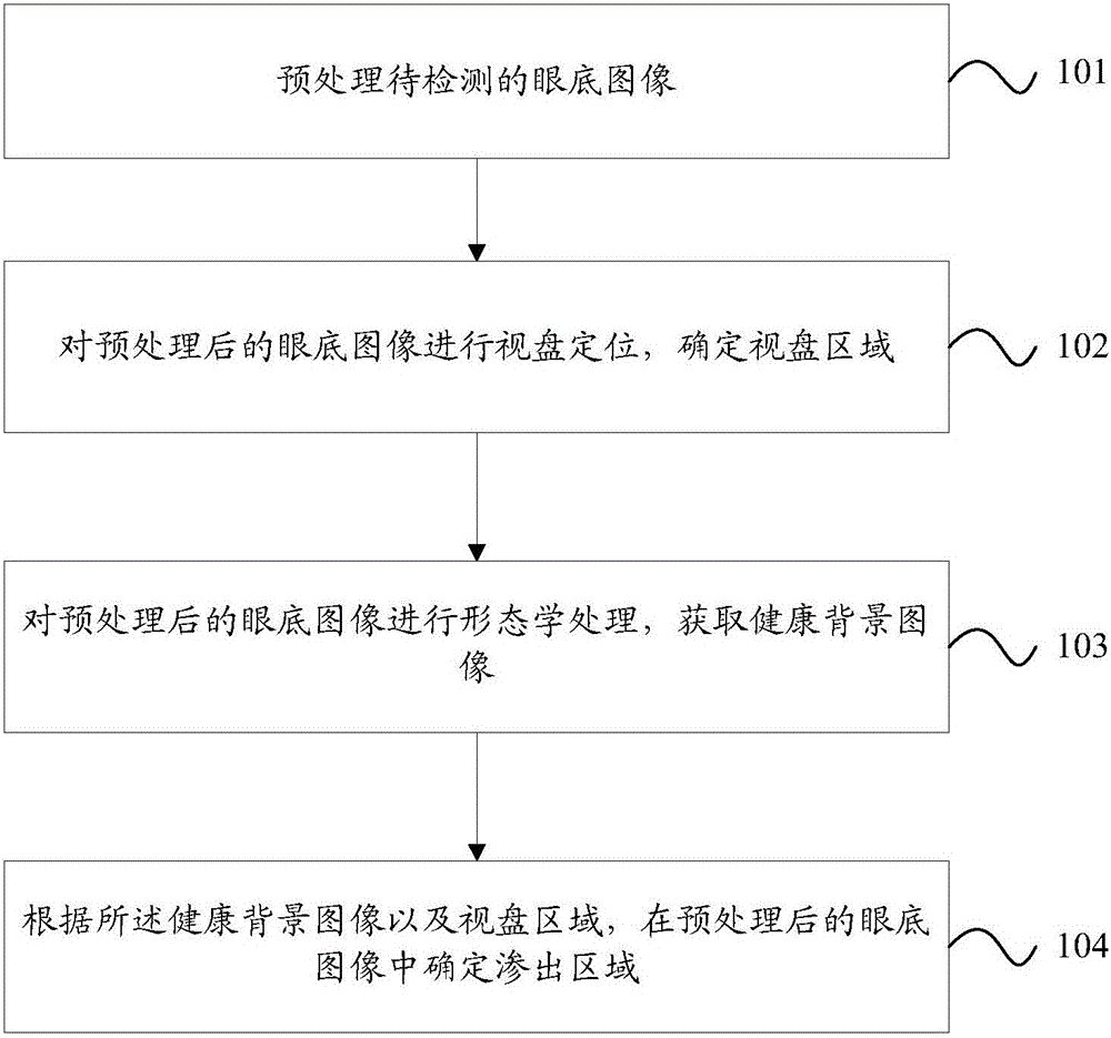
[0063] figure 1 It is a flow chart of the fundus image lesion detection method provided by Embodiment 1 of the present invention. Such as figure 1 As shown, the fundus image lesion detection method provided in this embodiment includes the following steps:

[0064] Step 101: Preprocessing the fundus image to be detected.

[0065] Wherein, step 101 ...


PUM


Abstract

The application discloses a lesion detection method and apparatus of an eye fundus image. The method comprises the following steps: carrying out pretreatment on a to-be-detected eye fundus image; carrying out optic disk positioning on the eye fundus image after pretreatment and determining an optic disk region; carrying out morphological processing on the eye fundus image after pretreatment to obtain a healthy background image; and according to the healthy background image and the optic disk region, determining an effusion region in the eye fundus image after pretreatment. Therefore, the detection speed of the eye fundus image can be increased and accuracy of positioning of a lesion region can be improved.

Description

technical field [0001] The invention relates to the field of computer vision information, in particular to a method and device for detecting fundus image lesions. Background technique [0002] With the development of computer graphics processing technology, the analysis of fundus images no longer depends solely on ophthalmologists' naked eyes. As early as the 1970s and 1980s, foreign scholars had proposed the technology of automatic detection of hard exudation and hemorrhage based on fundus image processing and analysis, and conducted a lot of research. Relying on computer graphics processing technology to quickly and accurately identify lesions in fundus images can effectively solve the problem of lack of quantitative means in traditional manual qualitative analysis, and also save a lot of manpower, material resources and time, providing a great opportunity for the implementation of large-scale screening of lesions basic condition. The research results have great practica...

Claims


Application Information

Patent Timeline
Patent Type & Authority Applications(China)
IPC IPC(8): G06T7/00G06T7/12G06T7/136
CPCG06T7/0012G06T2207/10024G06T2207/30041
Inventor 贾霞魏然盛斌殷本俊
Owner NANJING ZHONGXING XIN SOFTWARE CO LTD
Why Eureka
Try Eureka