Pulmonary nodule diagnosis method based on double-mode extreme learning machine

A technology of extreme learning machine and pulmonary nodules, which is applied in radiological diagnostic image/data processing, computer components, character and pattern recognition, etc., can solve problems such as unsupervised, unfavorable classification, misdiagnosis, etc. The method is simple and easy to implement Effect

Active Publication Date: 2017-09-22
TAIYUAN UNIV OF TECH
View PDF4 Cites 4 Cited by
  • Summary
  • Abstract
  • Description
  • Claims
  • Application Information

AI Technical Summary

Problems solved by technology

[0002] In the existing aided diagnosis system for pulmonary nodules, the classification of benign and malignant nodules is mainly based on single-modal image features (such as CT, MRI). Relying on a single feature will bring the risk of misdiagnosis
[0003] In the existing self-encoder network, the initial weights are randomly generated and unsupervised learning is adopted, the resulting image features are not very distinguishable, which is not conducive to classification

Method used


Image

  • Pulmonary nodule diagnosis method based on double-mode extreme learning machine
  • Pulmonary nodule diagnosis method based on double-mode extreme learning machine
  • Pulmonary nodule diagnosis method based on double-mode extreme learning machine

Examples


Embodiment Construction

[0056] The present invention will be described in detail below in conjunction with specific embodiments.

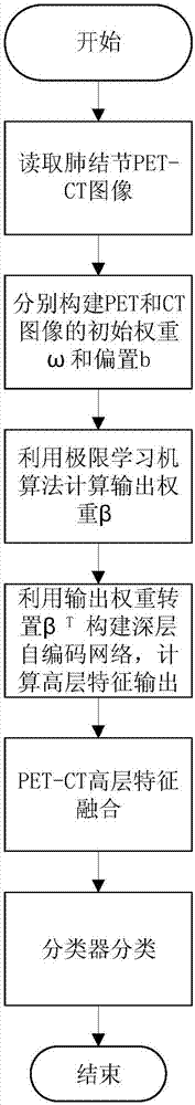
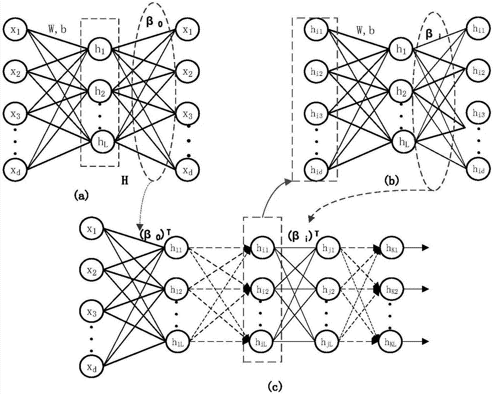
[0057] The deep extreme learning machine self-encoding is composed of N-layer networks, including an input layer and N-1 hidden layers, and the number of neurons in the input layer depends on the size of the input image. In the present invention, the linear interpolation method is used to convert The uniform size of PET-CT images in the dataset is 30×30, so the number of neurons in the input layer is 900. The number of neurons in the hidden layer can be given randomly. In the present invention, 1800 neurons are set in the hidden layer, and the Sigmoid function is used as the activation function of the hidden layer. For images of different modalities, the number of layers of the entire deep network is also different. The number of deep network layers corresponding to CT images is 7 layers, and the number of deep network layers corresponding to PET images is 5 layers.

[0...


PUM


Abstract

The invention discloses a pulmonary nodule diagnosis method based on a double-mode extreme learning machine. The method comprises the steps that firstly, pulmonary nodule PET-CT images are subjected to autonomous characteristic learning by using a self-encoding network with the supervision depth respectively, then, the extracted CT characteristics and PET characteristics are subjected to characteristics layer fusion, and finally, the fusion characteristics are classified by using a classifier. The method is simple and easy to implement, the identification accuracy is high, and according to the PET-CT images of pulmonary nodules, the benign and malignancy of the pulmonary nodules in the PET-CT images can be automatically and accurately identified based on the deep learning technology and the characteristic fusion method respectively.

Description

technical field [0001] The invention relates to the diagnosis of pulmonary nodules, in particular to a method for diagnosing pulmonary nodules based on a dual-mode extreme learning machine. Background technique [0002] In the existing auxiliary diagnosis system for pulmonary nodules, the classification of benign and malignant nodules is mainly based on single modality image features (such as CT, MRI). Relying on a single feature will bring the risk of misdiagnosis. [0003] In the existing self-encoding network, the initial weights are randomly generated and unsupervised learning is adopted, and the resulting image features are not very distinguishable, which is not conducive to classification. [0004] In order to solve the above problems, in the present invention, PET-CT images are used to diagnose pulmonary nodules by combining two modal image features, and the autoencoding algorithm is optimized by using restricted difference and supervised encoding methods in the autoe...

Claims


Application Information

Patent Timeline
Patent Type & Authority Applications(China)
IPC IPC(8): A61B6/03G06K9/62
CPCA61B6/037A61B6/52G06V2201/031G06F18/214G06F18/24G06F18/253
Inventor 强彦葛磊赵涓涓马瑞青王华强梓林唐笑先杜晓平
Owner TAIYUAN UNIV OF TECH
Why Eureka
Try Eureka