Ultrasonic image de-noising and enhancement method

An ultrasound image and image technology, applied in the field of medical image processing, can solve the problems of not making full use of the directional advantages of Curvelet transformation, and the lack of ultrasound images, etc., to achieve the effects of clinical diagnosis, noise suppression, and enhancement of anatomical information

Active Publication Date: 2017-09-26
VINNO TECH (SUZHOU) CO LTD +1
View PDF2 Cites 2 Cited by
  • Summary
  • Abstract
  • Description
  • Claims
  • Application Information

AI Technical Summary

Problems solved by technology

This type of method does not make full use of the directional advantages of the Curvelet tr

Method used


Image

  • Ultrasonic image de-noising and enhancement method
  • Ultrasonic image de-noising and enhancement method
  • Ultrasonic image de-noising and enhancement method

Examples


Embodiment Construction

[0036] The present invention will be further described through examples below, so as to better understand the technical solution of the present invention. Proceed as follows:

[0037] 1. Use Curvelet transform to decompose the image, the number of selected scales is 6 (including low-frequency subbands), and for the second coarsest scale level, the number of selected directions is 16.

[0038] 2. Multiply the low-frequency sub-band coefficients by a small gain coefficient, which is 0.8 in the embodiment.

[0039]

[0040] C 1,1 (i,j) are the original low frequency subband coefficients, are the processed low-frequency subband coefficients.

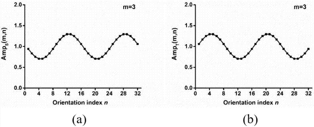
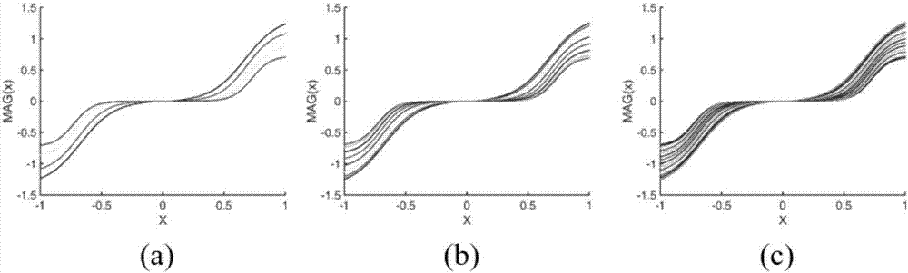
[0041] 3. Using a direction-selective high-frequency enhancement function MAG m,n (x), processing the coefficients of each high-frequency subband of Curvelet, which is defined as follows:

[0042] MAG m,n (x) = Amp S (m,n)*a*sigm(c(x-T(m,n)-b))-sigm(-c(x-T(m,n)+b))

[0043] in,

[0044] x=abs(C m,n (i, j)), C m,n (i, j) are ...


PUM


Abstract

The invention relates to the image processing field and discloses an ultrasonic image de-noising and enhancement algorithm. The ultrasonic image de-noising and enhancement method comprises steps of (1) using Curvelet transformation to perform decomposition on an image to obtain a low frequency sub-band and high frequency sub-band of different sizes and different directions, (2) multiplying a low frequency sub-band coefficient with a small gain coefficient and inhibiting image background information, (3) adopting a high frequency gain function having a direction selection to process a Curvelet high frequency sub-band coefficient, configuring a big gain multiple and a small threshold for the high frequency sub-band close to a horizontal direction, configuring a small gain multiple and a big threshold for the high frequency sub-band close to a perpendicular direction, and (4) performing Curvelet inverse transformation on the processed coefficient to obtain an enhanced image. The ultrasonic image de-noising and enhancement method can effectively remove spot noise of the ultrasonic image and can enhance edge information in the image.

Description

technical field [0001] The invention relates to an ultrasonic image denoising and enhancement method, which belongs to the field of medical image processing. Background technique [0002] Medical ultrasound imaging technology has been widely used in medical auxiliary diagnosis because of its non-invasive, non-radiation, real-time and high cost performance. However, due to the inherent mechanism of imaging in medical ultrasound images, there are a lot of speckle noise in the image, which affects the image quality. Severe speckle noise will cover up the detailed information of the image, which will interfere with the subsequent image feature extraction and recognition, quantitative analysis and clinical diagnosis. In addition, the anatomical information of ultrasound images is insufficient, the contrast is not good, and the lesion tissue is not easy to distinguish, which also greatly affects the accuracy of clinical diagnosis. Therefore, suppressing the speckle noise of ultr...

Claims


Application Information

Patent Timeline
IPC IPC(8): G06T5/00G06T5/10G06T5/20
CPCG06T5/002G06T5/10G06T5/20G06T2207/10132G06T2207/20048
Inventor 李豪张诗杰马睿陈惠人奚水张珏方竞
Owner VINNO TECH (SUZHOU) CO LTD
Why Eureka
Try Eureka