

Application of TNFR2
A kind of use and drug technology, applied in the field of TNFR2 as the target, can solve the problem of inability to save cardiomyocytes, and achieve the effects of repairing, inhibiting excessive differentiation, and enhancing expression and activity
- Summary
- Abstract
- Description
- Claims
- Application Information
AI Technical Summary
Problems solved by technology
Method used
Image

Examples
Embodiment 1
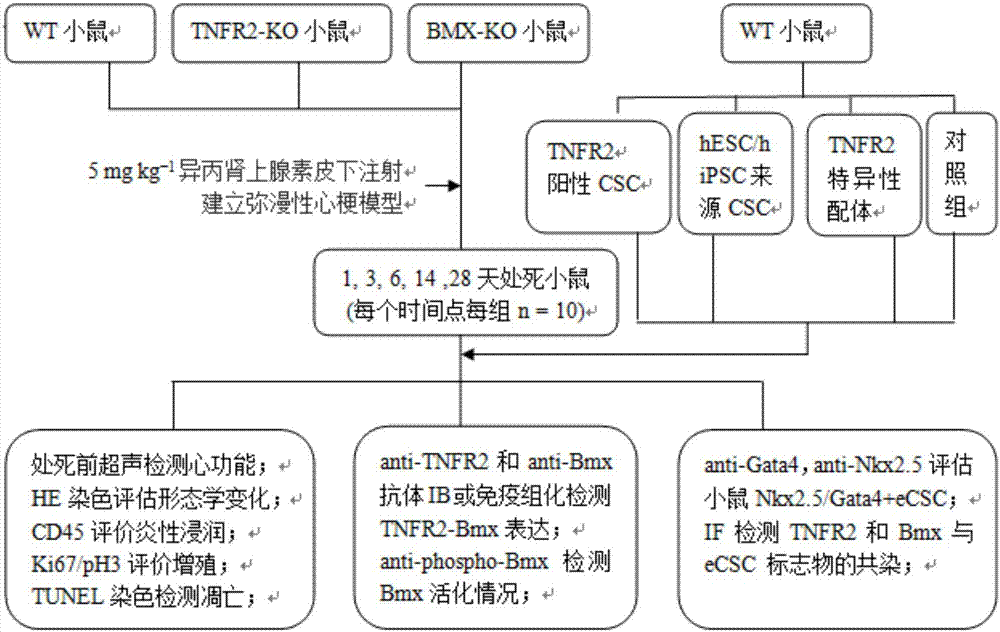
[0030] Embodiment 1: establish myocardial infarction model
[0031] All animal experiments in this example were approved by the Animal Ethics Committee.
[0032] experimental method:
[0033] 1) Coronary artery ligation: After anesthetizing the mouse, guide a flexible tube from the oral cavity into the trachea to connect to the ventilator, make an oblique incision on the left chest from the lower right to the upper left, and use a scalpel along the lower rib in the fourth intercostal space The intercostal muscles were incised to enter the thoracic cavity, and the heart was gently squeezed out. The proximal end of the left anterior descending artery was quickly sutured between the left atrial appendage and the conus pulmonary artery, and the proximal end of the left anterior descending branch was quickly sutured. At the same time, the changes of the electrocardiogram were observed. The syringe draws air out of the chest cavity to restore negative chest pressure. Mice with eje...
Embodiment 2
[0035] Embodiment 2: TNFR2-Bmx expression increases in human ischemic heart
[0036] The experimental method of this embodiment is to use anti-TNFR2 and anti-Bmx antibodies to perform IB or immunohistochemical detection of the expression of TNF receptor molecules and downstream effector molecules. The experimental samples are paraffin-embedded tissue sections obtained from Dr. Bradley of the University of Cambridge, UK during heart transplantation.
[0037] Its specific experimental method is:
[0038] 1) Immunofluorescence: tissue sections were fixed, Triton-X100 permeabilized, blocked with 1% BSA (bovine serum albumin) for 30 minutes, then rinsed with PBS (phosphate buffered saline), incubated with corresponding antibodies for 2 hours, rinsed with PBS, corresponding The fluorescent secondary antibody was incubated in the dark for 1 hour, the slide was mounted with glycerol after rinsing with PBS, observed and photographed under a fluorescent microscope.
[0039] 2) Western...
Embodiment 3
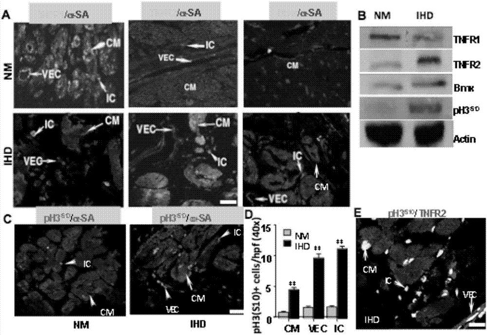
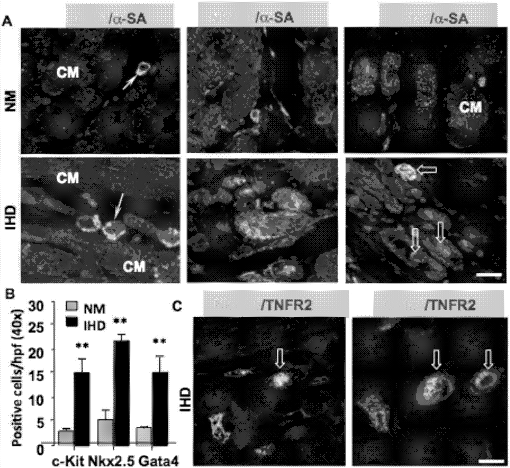
[0042] Example 3: TNFR2 is co-expressed with Nkx2.5+ / Gata4+CSC markers in ischemic heart
[0043] Wild-type, TNFR2-KO and Bmx-KO mice received a single subcutaneous injection of 5 mg kg-1 isoproterenol in the loose skin of the neck and were sacrificed at 1, 3, 6, 14 or 28 days, respectively (n=10 per group per time point). Cardiac function was detected by echocardiography before sacrifice. Use anti-TNFR2 and anti-Bmx antibodies for IB or immunohistochemical detection of TNFR2-Bmx expression. Bmx activation was detected by anti-phospho-Bmx. Mouse eCSCs were evaluated using anti-Gata4, anti-Nkx2.5. Simultaneously detect the expression of TNFR2 and Bmx and eCSC markers. Possible cardiac precursors were detected by immuno-double labeling for CSC markers (Gata4, NKx2.5 and α-SA).
[0044] The experimental results are attached figure 2 As shown in A, only some free cells are positive for Nkx2.5 and Gata4 in the normal heart. In the ischemic region of the ischemic heart, such...
PUM
 Abstract
Description
Claims
Application Information