Method and apparatus for automatically identifying a region of interest in three-dimensional CT image

A region of interest, CT image technology, applied in the field of medical image processing, can solve problems such as blurred boundaries and missed detections

Active Publication Date: 2017-12-15
HANGZHOU SHENRUI BOLIAN TECH CO LTD +1
View PDF6 Cites 27 Cited by
  • Summary
  • Abstract
  • Description
  • Claims
  • Application Information

AI Technical Summary

Problems solved by technology

Although CT images are widely used due to the advantages of clear images and high resolution, CT images also have the characteristics of displaying more tissues and blurred boundaries between tissues. In identifying the region of interest, there may be missed detection

Method used


Image

  • Method and apparatus for automatically identifying a region of interest in three-dimensional CT image
  • Method and apparatus for automatically identifying a region of interest in three-dimensional CT image
  • Method and apparatus for automatically identifying a region of interest in three-dimensional CT image

Examples


Embodiment Construction

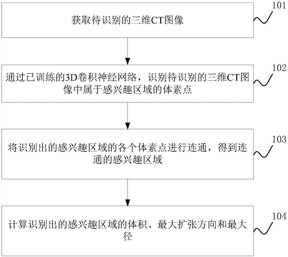
[0061] The method for identifying a region of interest proposed in the embodiment of the present application is applied to a CT image, with the purpose of automatically detecting a region of interest from a three-dimensional CT image, and improving the efficiency and accuracy of identifying the region of interest.

[0062] The CT image mentioned in the embodiment of the present application is an image of a certain part or an organ of a human body obtained through CT scanning, for example, it may be a scanned image of a lung or a scanned image of a human skeleton.

[0063] The region of interest mentioned in the embodiment of the present application is the scanned lesion area of ​​a certain part of the human body, for example: if the scanned CT image of the human lung is obtained, the region of interest may be a pulmonary nodule region; or If the scan is a CT image of the kidney, the area of ​​interest may be the area of ​​kidney stones.

[0064] The method for identifying a re...


PUM


Abstract

The present invention discloses a method and an apparatus for automatically identifying a region of interest in a three-dimensional CT image. The method includes: training a 3D convolution neural network through a three-dimensional CT image marked with a region of interest to obtain a 3D convolutional neural network with a high identification rate for the region of interest; then, through the trained 3D convolution neural network, identifying the obtained three-dimensional CT image and obtaining the region of interest; and calculating the volume, the expansion direction and the expansion diameter of the interest of interest. With the method of the embodiments in the invention, the region of interest representing the lesion can be obtained automatically and efficiently, and the identifying accuracy is also higher. In addition, the volume, the expansion direction and the expansion diameter of the lesion can be automatically calculated, therefore, helping the doctor to make a more accurate judgment of the condition of a patient.

Description

technical field [0001] The invention relates to the field of medical image processing, in particular to a method and device for identifying a region of interest in a three-dimensional CT image. Background technique [0002] With the development of digital medical technology, the role of medical images in the medical diagnosis process has become more and more obvious, and it has gradually become the main tool for doctors to observe human lesions. When diagnosing a disease, doctors generally search for regions of interest representing lesions from medical images artificially based on experience. [0003] CT image is one of the most commonly used medical images. CT (full name in English: Computed Tomography, full name in Chinese: computerized tomography) images use precise and straight x-ray beams, gamma rays, ultrasound, etc., and are surrounded by highly sensitive detectors. In a certain part of the human body, it is obtained as one-by-one cross-sectional scanning. Although...

Claims


Application Information

Patent Timeline
Patent Type & Authority Applications(China)
IPC IPC(8): G06K9/32G06T7/00G06T7/62
CPCG06T7/0012G06T7/62G06T2207/10081G06T2200/04G06T2207/20081G06T2207/20084G06V10/25G06V2201/03
Inventor 周振张番栋庞亮张树孔明王洪振何哲琪李一鸣
Owner HANGZHOU SHENRUI BOLIAN TECH CO LTD
Why Eureka
Try Eureka