Automatic identification and statistics method for white blood cells in gynecologic microscopic image

A microscopic image and automatic recognition technology, applied in image enhancement, image analysis, image data processing, etc., can solve problems such as slow convergence, slow detection speed, and failure to meet clinical requirements, so as to avoid the influence of subjective factors and save manpower The effect of material resources, time-saving and labor-saving technical means

Active Publication Date: 2017-12-19
青岛华晶生物技术有限公司
View PDF10 Cites 19 Cited by
  • Summary
  • Abstract
  • Description
  • Claims
  • Application Information

AI Technical Summary

Problems solved by technology

[0009] Li Wenhui of Nanchang Hangkong University published a paper "Key Technology Research on Microscopic Image Recognition of Saline Leucorrhea Cells" in 2013, using the level set active contour model segmentation method to segment white blood cells. This model has good segmentation results for weak edges, but The convergence is slow. If there are multiple white blood cells in an image, the detection speed is slow and cannot meet the clinical requirements.

Method used


Image

  • Automatic identification and statistics method for white blood cells in gynecologic microscopic image
  • Automatic identification and statistics method for white blood cells in gynecologic microscopic image
  • Automatic identification and statistics method for white blood cells in gynecologic microscopic image

Examples


specific Embodiment approach

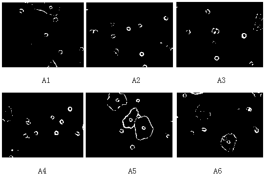
[0043] The specific embodiments of the present invention are as follows: a method for automatic identification and statistics of white blood cells in gynecological microscopic images, comprising the following steps:

[0044] Step 1, grayscale the color image (such as figure 2 shown);

[0045] Step 2, using image segmentation technology to obtain the mask image of white blood cells:

[0046] The first step: use the Sobel operator to perform edge detection, add the image after edge detection to the grayscale image, and realize the edge enhancement of each component to be detected in the image;

[0047] The second step: use the Otsu method to operate the image to obtain a binarized image;

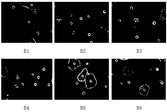
[0048] Step 3: Invert the mask image of the epithelial cells:

[0049] The mask image acquisition steps of epithelial cells are as follows:

[0050] The first step: use the Sobel operator to perform edge detection, add the image after edge detection to the grayscale image, and realize the...


PUM


Abstract

The invention discloses an automatic identification and statistics method for white blood cells in a gynecologic microscopic image. A color image is firstly converted into a gray image, and an image segmentation method is used to obtain a template for the white blood cells; AND-operation is carried out on the template and the gray image; each small area is segmented; and by using the roundness of the white blood cells, the length-width ratio of an external rectangle, the area and the ratio of a cell nucleus to the cell, identification on the white blood cells is realized. Automatic identification and statistics are realized, clinical requirements are met, artificial detection and cell identification by using cell staining are replaced, and the manpower and the material resources are saved greatly.

Description

technical field [0001] The invention relates to a method for automatically identifying and counting white blood cells in gynecological microscopic images. Background technique [0002] Cell image processing is a typical application of digital image processing technology in the medical field. However, due to the complexity of cell images, currently it mainly relies on manual reading, and doctors observe with naked eyes to find some diseased cells from a large number of cells. Long-term high-intensity observation work is very easy to make people tired, and requires doctors to have rich clinical experience and comprehensive pathological analysis expertise. The accuracy and reliability of diagnostic results are affected by the subjective factors of doctors. Due to inconsistent observation conditions and inevitable systematic errors, the final diagnosis results often show a high rate of misdiagnosis. [0003] To improve the above-mentioned problems, in addition to improving the ...

Claims


Application Information

Patent Timeline
Patent Type & Authority Applications(China)
IPC IPC(8): G06T7/00G06T7/12G06T7/136G06T7/62
CPCG06T7/0012G06T2207/10061G06T2207/30024G06T2207/30242G06T7/12G06T7/136G06T7/62
Inventor 王训琨李金平秦丽
Owner 青岛华晶生物技术有限公司
Why Eureka
Try Eureka