Ultrasound image processing method and device, ultrasonic diagnosis device and storage medium

A technology of image processing and ultrasound, applied in ultrasound/sonic/infrasonic image/data processing, ultrasound/sonic/infrasonic diagnosis, structure of ultrasound/sonic/infrasonic diagnostic equipment, etc., which can solve the problem of low accuracy of amniotic fluid detection results and other issues to achieve the effect of reducing misdiagnosis and improving accuracy

Active Publication Date: 2017-12-29
SONOSCAPE MEDICAL CORP
View PDF8 Cites 8 Cited by
  • Summary
  • Abstract
  • Description
  • Claims
  • Application Information

AI Technical Summary

Problems solved by technology

[0007] The invention provides an ultrasonic image processing method and device, an ultrasonic diagnostic device and a computer-readable storage medium, which are used to solve the problem of low accuracy of the existing amniotic fluid detection results

Method used


Image

  • Ultrasound image processing method and device, ultrasonic diagnosis device and storage medium
  • Ultrasound image processing method and device, ultrasonic diagnosis device and storage medium
  • Ultrasound image processing method and device, ultrasonic diagnosis device and storage medium

Examples


Embodiment Construction

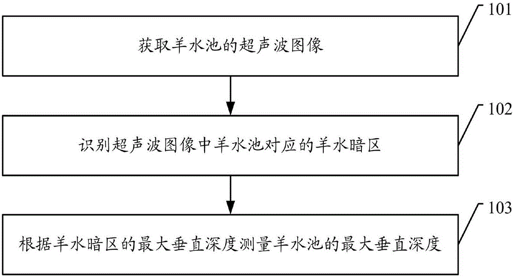
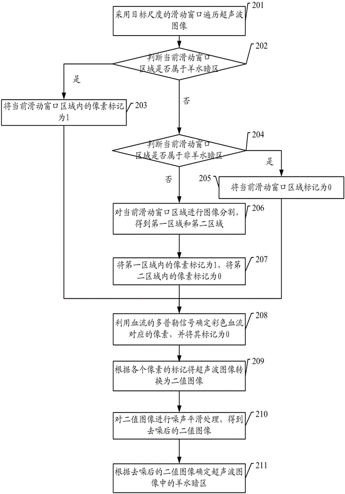
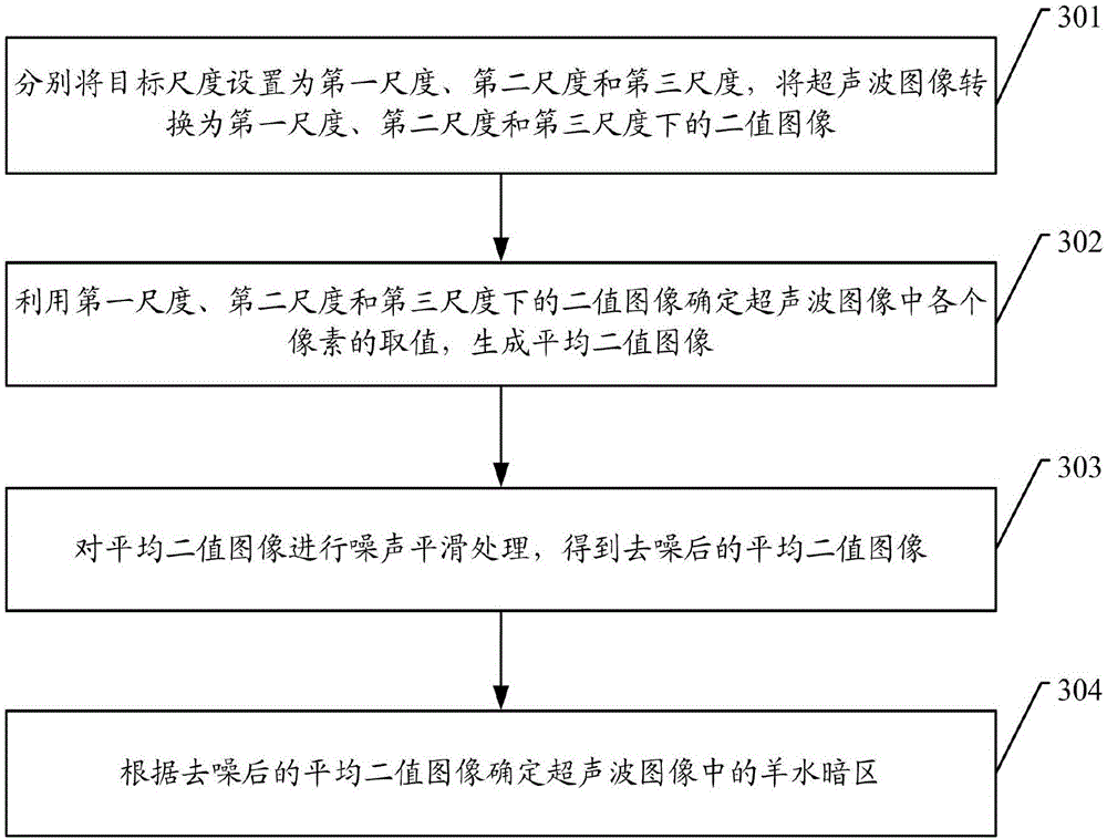
[0069] Embodiments of the present invention provide an ultrasonic image processing method and device, an ultrasonic diagnostic device, and a computer-readable storage medium, which are used to reduce human factors in the amniotic fluid measurement process and improve the accuracy of amniotic fluid detection results.

[0070] In order to enable those skilled in the art to better understand the solutions of the present invention, the following will clearly and completely describe the technical solutions in the embodiments of the present invention in conjunction with the drawings in the embodiments of the present invention. Obviously, the described embodiments are only It is an embodiment of a part of the present invention, but not all embodiments. Based on the embodiments of the present invention, all other embodiments obtained by persons of ordinary skill in the art without making creative efforts shall fall within the protection scope of the present invention.

[0071] The ter...


PUM


Abstract

The embodiment of the invention discloses an ultrasound image processing method and device, an ultrasonic diagnosis device and a computer readable storage medium, and relates to the field of image processing. The ultrasound image processing method and device, the ultrasonic diagnosis device and the computer readable storage medium are used for avoiding an impact which human factors have on a measurement result of the amniotic fluid volume as much as possible and improving the correctness of an amniotic fluid detection result and are beneficial to misdiagnosis reduction. The method comprises the steps that an ultrasound image of an amniotic fluid pocket is obtained; an amniotic fluid opaque area corresponding to the amniotic fluid pocket in the ultrasound image is identified; the largest vertical depth of the amniotic fluid pocket is measured according to the largest vertical depth of the amniotic fluid opaque area.

Description

technical field [0001] The invention relates to the field of ultrasonic diagnostic equipment, in particular to an ultrasonic image processing method and device, an ultrasonic diagnostic device, and a computer-readable storage medium. Background technique [0002] Abnormal amniotic fluid volume is closely related to fetal development and has become an important detection content during pregnancy. Due to the limitation of the examination time, the general ultrasound radiation time to the fetus should not be too long and the professional level and the function of the ultrasound equipment are limited. At present, it is not possible to diagnose various deformities in a short period of time for each pregnant woman in a single examination. Therefore, abnormal amniotic fluid volume becomes a prompt to find high-risk situations, and its detection and judgment are particularly important. It can prompt the testing doctor to conduct detailed examinations on the fetus in time. [0003] ...

Claims


Application Information

Patent Timeline
Patent Type & Authority Applications(China)
IPC IPC(8): A61B8/08A61B8/00G06T5/00G06T5/30G06T7/11
CPCA61B8/0833A61B8/44A61B8/488A61B8/5215A61B8/5269G06T5/002G06T5/30G06T7/11G06T2207/10136
Inventor 廖静秋黄子殷曹锡华许龙
Owner SONOSCAPE MEDICAL CORP
Why Eureka
Try Eureka