Tissue-derived exosome extraction method and application thereof

An extraction method and exosome technology, which are applied in the field of tissue-derived exosome extraction to achieve the effect of convenient operation and field expansion

Inactive Publication Date: 2017-12-29
SHANGHAI TONGJI HOSPITAL
View PDF12 Cites 15 Cited by
  • Summary
  • Abstract
  • Description
  • Claims
  • Application Information

AI Technical Summary

Problems solved by technology

However, there are no reports on how to process tissue samples and what method to use to extract tissue exosomes.

Method used


Image

  • Tissue-derived exosome extraction method and application thereof
  • Tissue-derived exosome extraction method and application thereof
  • Tissue-derived exosome extraction method and application thereof

Examples


Embodiment 1

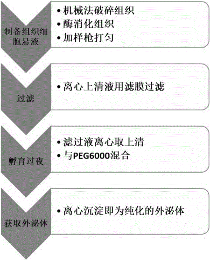
[0035] 1. Place the fresh tissue block in 350μl 1640 medium pre-cooled;

[0036] 2. Mince the tissue block into 1mm pieces with a sharp blade 3 size, operated on ice;

[0037] 3. Add 350 μl of 1 mg / ml collagenase IV and 2 μl of 0.2% (w / v) Dnase I, cut off the sharp part of the pipette tip, and gently pipette the mixture;

[0038] 4. Digest the above mixture on a constant temperature shaker at 37°C and 100 rpm for 90 minutes until the tissue pieces are invisible to the naked eye, then stop the digestion, and collect the samples at 4°C to delay their digestion;

[0039] 5. After all the tissue pieces are completely digested, transfer to a test tube, centrifuge at 3000g 4°C for 30min, absorb the supernatant, transfer to a new centrifuge tube, centrifuge at 13000g 4°C for 10min, and harvest the supernatant;

[0040] 6. Pass the supernatant through a 0.22μm filter, transfer the filtrate to a new centrifuge tube, centrifuge at 13,000g at 4°C for 10min, and collect the supernatant;...

Embodiment 2

[0049] 1. Place the cryogenically frozen (-80°C) tissue block in 350 μl 1640 medium pre-cooled (frozen tissue needs to be quickly placed in a 37°C water bath for 5 minutes);

[0050] 2. Mince the tissue block into 1mm pieces with a sharp blade 3 size, operated on ice;

[0051] 3. Add 350 μl of 1 mg / ml collagenase IV and 2 μl of 0.2% (w / v) Dnase I, cut off the sharp part of the pipette tip, and gently pipette the mixture;

[0052] 4. Digest the above mixture on a constant temperature shaker at 37°C and 100 rpm for 80 minutes until the tissue pieces are invisible to the naked eye, then stop the digestion, and collect the samples at 4°C to delay their digestion;

[0053] 5. After all the tissue pieces are completely digested, transfer to a test tube, centrifuge at 3000g 4°C for 30min, absorb the supernatant, transfer to a new centrifuge tube, centrifuge at 13000g 4°C for 10min, and harvest the supernatant;

[0054] 6. Pass the supernatant through a 0.22μm filter, transfer the fil...

Embodiment 3

[0058] 1. Place cryogenically frozen (liquid nitrogen) tissue pieces in pre-cooled 350 μl 1640 medium (frozen tissues need to be quickly placed in a 37°C water bath for 5 minutes);

[0059] 2. Mince the tissue block into 1mm pieces with a sharp blade 3 size, operated on ice;

[0060] 3. Add 350 μl of 1 mg / ml collagenase IV and 2 μl of 0.2% (w / v) Dnase I, cut off the sharp part of the pipette tip, and gently pipette the mixture;

[0061] 4. Digest the above mixture on a constant temperature shaker at 37°C and 100rpm for 100 minutes until the tissue pieces are invisible to the naked eye, then stop the digestion, and collect the samples at 4°C to delay their digestion;

[0062] 5. After all the tissue pieces are completely digested, transfer to a test tube, centrifuge at 3000g 4°C for 30min, absorb the supernatant, transfer to a new centrifuge tube, centrifuge at 13000g 4°C for 10min, and harvest the supernatant;

[0063] 6. Pass the supernatant through a 0.22μm filter, transfe...


PUM

PropertyMeasurementUnit
pore sizeaaaaaaaaaa
diameteraaaaaaaaaa

Abstract

The invention relates to a tissue-derived exosome extraction method and belongs to the technical field of biology. The tissue-derived exosome extraction method is used for extracting exosomes directly based on enzymatic lysis and includes the steps of firstly, cutting tissues mechanically and adding collagenase and Dnase for lysis; secondly, subjecting lysed mixed liquor to exosome operation by a PEG method. The tissue-derived exosome extraction method has the advantages that the tissue-derived exosome extraction method obtains the exosomes from the tissues directly for further studying, overcomes the difficulty in extracting the exosomes from the tissues directly and expands the field of exosome researches by extracting the exosomes by means of enzymolysis-filtration-centrifugation, thereby being of great significance to promoting the exosome researches.

Description

technical field [0001] The invention relates to the field of biotechnology, in particular to a method for extracting tissue-derived exosomes and its application. Background technique [0002] Exosomes (exosomes) are vesicle bodies secreted by a variety of living cells, with a diameter of 50-150 nm. They are produced after the fusion of multivesicular membranes and cell membranes, and contain various components such as proteins and RNA. Subsequent studies have found that exosomes can carry RNA for the exchange of intercellular substances, and can perform biological functions in recipient cells, which means that exosomes are a new way for the exchange of genetic material between cells, pioneering the development of genetics. new mechanism on . The latest research shows that exosomes are associated with cancer metastasis, and cancer cells create a microenvironment suitable for their survival at the far end through macromolecules such as exosome transport proteins and RNA. Tum...

Claims


Application Information

Patent Timeline
Patent Type & Authority Applications(China)
IPC IPC(8): C12N5/071
CPCC12N5/0602C12N2509/00
Inventor 李冬潘星昊权文强吴军录
Owner SHANGHAI TONGJI HOSPITAL
Why Eureka
Try Eureka