Ultrasound image processing method and system and ultrasound diagnosis equipment

A technology of ultrasonic image and processing method, which is applied in the directions of ultrasonic/sonic/infrasonic image/data processing, image data processing, ultrasonic/sonic/infrasonic diagnosis, etc. It can solve the problems of cumbersome operation steps and low efficiency, and improve the accuracy Accuracy and efficiency, direct and rapid identification and acquisition

Inactive Publication Date: 2018-01-12
SONOSCAPE MEDICAL CORP
View PDF4 Cites 27 Cited by
  • Summary
  • Abstract
  • Description
  • Claims
  • Application Information

AI Technical Summary

Problems solved by technology

The operation steps are cumbersome and the efficiency is low

Method used


Image

  • Ultrasound image processing method and system and ultrasound diagnosis equipment
  • Ultrasound image processing method and system and ultrasound diagnosis equipment
  • Ultrasound image processing method and system and ultrasound diagnosis equipment

Examples


Embodiment 1

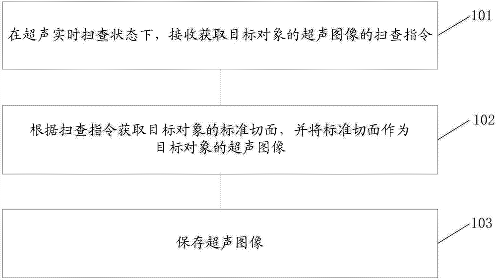
[0051] The first embodiment of the present invention will describe an ultrasonic image processing method in detail. The ultrasonic image processing method in this embodiment includes the following steps, as figure 1 as shown, figure 1 It is a flow chart of the ultrasonic image processing method described in the first embodiment of the present invention, an ultrasonic image processing method comprising the following steps:

[0052] 101. In the ultrasonic real-time scanning state, receive a scanning instruction for acquiring the ultrasonic image of the target object;

[0053] In the ultrasonic real-time scanning state, a scanning instruction input by a user to obtain an ultrasonic image of a target object is received. In this embodiment, the target object may be the tissues and organs to be examined, such as fetus, thyroid gland, breast, liver and so on. The scanning instructions may be scanning items of different pregnancy periods, such as cervical canal sagittal section, tha...

Embodiment 2

[0096] The second embodiment of the present invention describes an ultrasonic image processing method. The ultrasonic image processing method described in this embodiment includes the following steps, such as Figure 5 as shown, Figure 5 It is the flowchart of the ultrasonic image processing method described in the second embodiment of the present invention:

[0097] 501. In the ultrasonic real-time scanning state, receive a scanning instruction for acquiring an ultrasonic image of a target object;

[0098] 502. Obtain a standard slice of the target object according to the scan instruction, and use the standard slice as the ultrasonic image of the target object;

[0099] 503. Save the ultrasound image;

[0100] 504. Measure the standard section according to the preset measurement requirement, and obtain the measurement result.

[0101] This embodiment is further obtained on the basis of the first embodiment of the present invention. After the standard slice is obtained ac...

Embodiment 3

[0104] The third embodiment of the present invention will describe an ultrasonic image processing device in detail. For the specific structure of the ultrasonic image processing device, please refer to Image 6 , Image 6 It is a schematic diagram of the structure of the ultrasonic image processing device according to the third embodiment of the present invention. The ultrasonic image processing device includes an instruction receiving unit 601 , an image acquiring unit 602 and a storage unit 603 .

[0105] The instruction receiving unit 601 is configured to receive a scanning instruction for acquiring an ultrasonic image of a target object in a real-time ultrasonic scanning state.

[0106] The image acquiring unit 602 is configured to acquire a standard slice of the target object according to the scan instruction, and use the standard slice as an ultrasound image of the target object.

[0107] The storage unit 603 is configured to save the ultrasound image.

[0108] In the ...


PUM


Abstract

The invention discloses an ultrasound image processing method. In the ultrasound real-time scanning state, a scanning instruction of an ultrasound image of a target object is received and acquired; according to the scanning instruction, a standard section of the target object is acquired and regarded as the ultrasound image of the target object; the ultrasound image is stored. The invention further discloses an ultrasound image processing device and ultrasound diagnosis equipment. According to the ultrasound image processing method and system and the ultrasound diagnosis equipment, in the ultrasound real-time scanning state, the standard section of the target object is acquired and regarded as the ultrasound image of the target object according to the scanning instruction, direct fast recognition and acquirement of the standard section are achieved, and manual freezing or thawing operation is not needed, so that the accuracy and efficiency of scanning are improved.

Description

technical field [0001] The present invention relates to the technical field of ultrasonic diagnostic equipment, in particular to an ultrasonic image processing method and system, and ultrasonic diagnostic equipment. Background technique [0002] At present, the advantageous medical resources are highly concentrated in big cities and affiliated hospitals of medical colleges. Ultrasound image scanning requires operators to have standardized operating techniques, rich clinical experience, and high technical threshold. However, there is a shortage of resources in grass-roots hospitals, and there are still many problems in the operation of the operator, such as irregular techniques and little clinical experience. [0003] Moreover, the existing scanning process is roughly as follows: After scanning a series of ultrasound images, freeze the images first, then play back the movie by sliding the trackball, etc., manually find the required standard section, then unfreeze the images, ...

Claims


Application Information

Patent Timeline
Patent Type & Authority Applications(China)
IPC IPC(8): A61B8/08A61B8/00G06K9/62G06T7/00
CPCA61B8/00A61B8/08A61B8/14G06T7/00G06F18/00
Inventor 李萍唐艳红廖静秋许龙
Owner SONOSCAPE MEDICAL CORP
Why Eureka
Try Eureka