Ultrasonic and CT/MR image fusion surgical navigation system and method based on optical localization rectification
An ultrasound imaging system and image fusion technology, applied in surgical navigation systems, surgery, medical science, etc., can solve the problems of soft tissue image drift, lack, and large positioning, and achieve the effect of reducing surgical navigation errors and accurate positioning.
- Summary
- Abstract
- Description
- Claims
- Application Information
AI Technical Summary
Problems solved by technology
Method used
Image
Examples
Embodiment 1
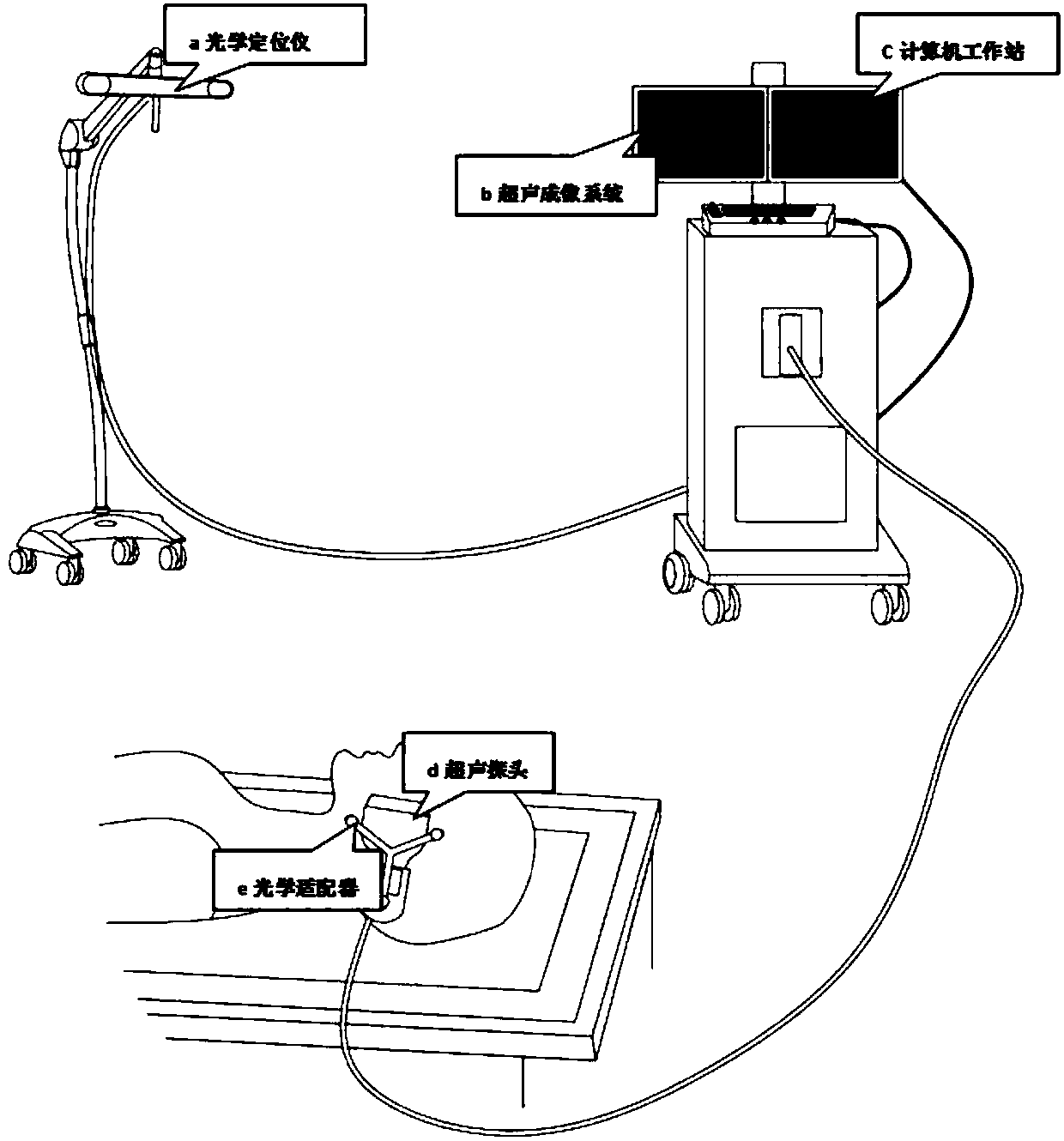
[0028] A surgical navigation system based on fusion of ultrasound and CT / MR images based on optical positioning, such as figure 1 As shown, it is used for real-time monitoring of soft tissue deformation in craniomaxillofacial surgery. The fusion system includes an ultrasonic imaging system, a tracking and positioning system, and a computer workstation. The ultrasonic imaging system includes an ultrasonic probe and an image workstation. The tracking and positioning system Including an adapter fixed on the ultrasonic probe and a locator for obtaining the coordinates of the ultrasonic probe according to the marker, the computer workstation records its spatial position according to the real-time ultrasonic image obtained by the ultrasonic probe, performs three-dimensional reconstruction, and displays the fused image .
[0029] Further, the ultrasonic probe is a two-dimensional ultrasonic probe.
[0030] Further, the imaging workstation is a portable color Doppler ultrasound diagn...
Embodiment 2
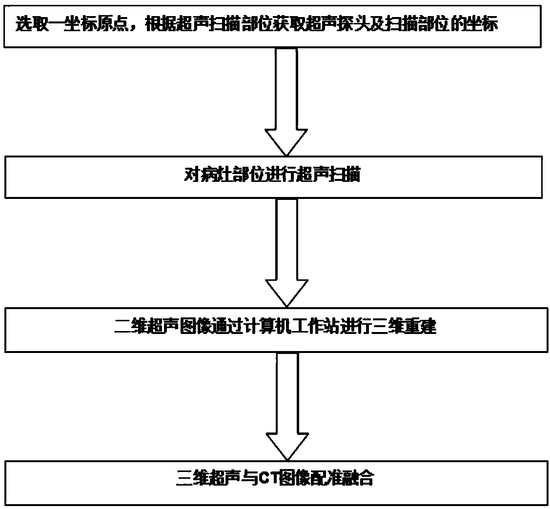
[0035] Apply the image fusion surgical navigation system of the present invention to carry out the method for real-time ultrasound and CT / MR image fusion, such as figure 2 shown, including the following steps:
[0036] The ultrasonic imaging system, the tracking and positioning system and the computer workstation are connected by communication;
[0037] Selecting a coordinate origin to obtain the position coordinates of the ultrasonic probe;
[0038] Ultrasound scan imaging of the ultrasound scan site;
[0039] Recording the spatial position information of the two-dimensional fan-shaped ultrasonic image according to the obtained position coordinates of the ultrasonic probe;
[0040] Performing three-dimensional reconstruction on the two-dimensional ultrasound image in the computer workstation;
[0041] The three-dimensional ultrasound image is registered and fused with the CT / MR image imported into the computer workstation, image 3An ultrasound and CT fusion image obtain...
Embodiment 3
[0043] In order to verify the feasibility of the above method and system, CT and free-style ultrasound scanning with optical positioning were performed on patients with facial foreign bodies and parotid gland tumors. The connected and integrated system was running normally under the joint on-site monitoring of oral and maxillofacial surgeons, sonographers, and engineers, and successfully obtained ultrasound images of the lesion area according to the image acquisition standards of the engineers.
[0044] Fusion image analysis:
[0045] In order to test the effect of image fusion, we imported the fused ultrasound and CT data into the image processing software MIPAV again, and superimposed the ultrasound and CT data. Then the effect of image fusion is analyzed through subjective and objective evaluation methods.
[0046] The subjective evaluation method is that two experienced sonographers and radiologists jointly review the original image and the fused image, and evaluate the f...
PUM
 Abstract
Description
Claims
Application Information
 - R&D
- Intellectual Property
- Life Sciences
- Materials
- Tech Scout
- Unparalleled Data Quality
- Higher Quality Content
- 60% Fewer Hallucinations
Browse by: Latest US Patents, China's latest patents, Technical Efficacy Thesaurus, Application Domain, Technology Topic, Popular Technical Reports.
© 2025 PatSnap. All rights reserved.Legal|Privacy policy|Modern Slavery Act Transparency Statement|Sitemap|About US| Contact US: help@patsnap.com


