Fluorescent dye labeling aptamer for immune globulin E with sensitive fluorescence anisotropy response
A nucleic acid aptamer and immunoglobulin technology, applied in the field of analysis and detection, can solve the problems of no obvious signal change, influence sensitivity, and insignificant fluorescence polarization, and achieve the effect of significant signal change, large signal change range and good affinity.
- Summary
- Abstract
- Description
- Claims
- Application Information
AI Technical Summary
Problems solved by technology
Method used
Image
Examples
Embodiment 1
[0065] Example 1: Detection of IgE using IgE37-T9-FAM fluorescence anisotropy (fluorescence polarization)
[0066] 1. Mix IgE37-T9-FAM, IgE and binding buffer to obtain a reaction system. The final concentration of IgE37-T9-FAM in the reaction system was 10 nM. The final concentrations of IgE in the reaction system were 0nM, 0.01nM, 0.02nM, 0.05nM, 0.1nM, 0.2nM, 0.5nM, 1nM, 2nM, 5nM, 10nM, 20nM, 50nM, 100nM and 200nM, respectively.
[0067] The above-mentioned IgE37-T9-FAM is an aptamer obtained by FAM-labeling the ninth nucleotide T of the nucleic acid aptamer IgE37.
[0068] 2. Incubate the reaction system in an ice box (4°C) for 20 minutes.
[0069] 3. The fluorescence anisotropy value (r) of IgE37-T9-FAM was measured at 25°C using a fluorometer.
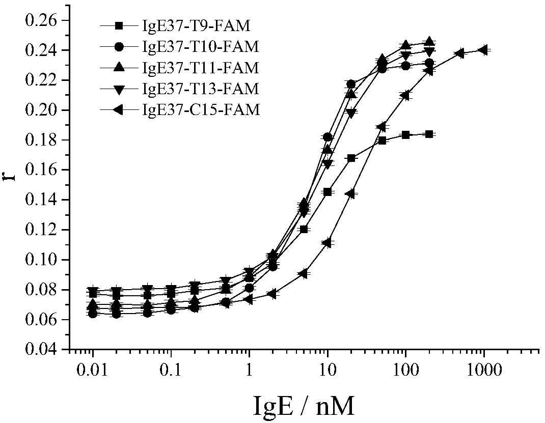
[0070] The result is as figure 1 shown. figure 1 It was shown that the fluorescence anisotropy of IgE37-T9-FAM increased gradually with the increase of IgE concentration. At high concentration of IgE, the fluorescence anisot...
Embodiment 2
[0071] Example 2: Detection of IgE using IgE37-T10-FAM fluorescence anisotropy (fluorescence polarization)
[0072] 1. Mix IgE37-T10-FAM, IgE and binding buffer to obtain a reaction system. The final concentration of IgE37-T10-FAM in the reaction system was 10 nM. The final concentrations of IgE in the reaction system were 0nM, 0.01nM, 0.02nM, 0.05nM, 0.1nM, 0.2nM, 0.5nM, 1nM, 2nM, 5nM, 10nM, 20nM, 50nM, 100nM and 200nM, respectively.
[0073] The above-mentioned IgE37-T10-FAM is an aptamer obtained by labeling the 10th nucleotide T of the nucleic acid aptamer IgE37 with FAM.
[0074] 2. Incubate the reaction system in an ice box (4°C) for 20 minutes.
[0075] 3. The fluorescence anisotropy value (r) of IgE37-T10-FAM was measured at 25°C with a fluorometer.
[0076] The result is as figure 1 shown. figure 1 It is shown that the fluorescence anisotropy of IgE37-T10-FAM increases gradually with the increase of IgE concentration. At high concentration of IgE, the fluorescenc...
Embodiment 3
[0077] Example 3: Detection of IgE using IgE37-T11-FAM fluorescence anisotropy (fluorescence polarization)
[0078] 1. Mix IgE37-T11-FAM, IgE and binding buffer to obtain a reaction system. The final concentration of IgE37-T11-FAM in the reaction system was 10 nM. The final concentrations of IgE in the reaction system were 0nM, 0.01nM, 0.02nM, 0.05nM, 0.1nM, 0.2nM, 0.5nM, 1nM, 2nM, 5nM, 10nM, 20nM, 50nM, 100nM and 200nM, respectively.
[0079] The above-mentioned IgE37-T11-FAM is an aptamer obtained by labeling the 11th nucleotide T of the nucleic acid aptamer IgE37 with FAM.
[0080] 2. Incubate the reaction system in an ice box (4°C) for 20 minutes.
[0081] 3. The fluorescence anisotropy value (r) of IgE37-T11-FAM was measured at 25°C with a fluorometer.
[0082] The result is as figure 1 shown. figure 1 It was shown that the fluorescence anisotropy of IgE37-T11-FAM increased gradually with the increase of IgE concentration. At high concentration of IgE, the fluorescen...
PUM
 Abstract
Description
Claims
Application Information
 - R&D
- Intellectual Property
- Life Sciences
- Materials
- Tech Scout
- Unparalleled Data Quality
- Higher Quality Content
- 60% Fewer Hallucinations
Browse by: Latest US Patents, China's latest patents, Technical Efficacy Thesaurus, Application Domain, Technology Topic, Popular Technical Reports.
© 2025 PatSnap. All rights reserved.Legal|Privacy policy|Modern Slavery Act Transparency Statement|Sitemap|About US| Contact US: help@patsnap.com


