Analyzing and processing method and device for tumor image

An image analysis and tumor technology, applied in the field of image processing, can solve the problem of not having the resolution information in the depth direction, and achieve the effect of comprehensive observation

Inactive Publication Date: 2018-05-04
SHENZHEN PEOPLES HOSPITAL
View PDF3 Cites 5 Cited by
  • Summary
  • Abstract
  • Description
  • Claims
  • Application Information

AI Technical Summary

Problems solved by technology

Moreover, the current near-infrared fluorescence imaging systems can only perform two-dimensional plane imaging, and do not have resolution information in the depth direction.

Method used


Image

  • Analyzing and processing method and device for tumor image
  • Analyzing and processing method and device for tumor image
  • Analyzing and processing method and device for tumor image

Examples


Embodiment approach

[0028] The tumor image analysis and processing method of the present application, one embodiment thereof, includes:

[0029] The stiffness information from ultrasound imaging is combined with the blood oxygen information provided by photoacoustic imaging to analyze tumor images.

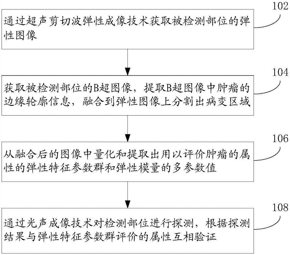
[0030] In one embodiment, as figure 1 As shown, the tumor image analysis and processing method of the present application may specifically include the following steps:

[0031] Step 102: Obtain an elastic image of the detected part by ultrasonic shear wave elastography.

[0032] Step 104: Acquire the B-ultrasound image of the detected part, extract the edge contour information of the tumor in the B-ultrasound image, and fuse it to the elastic image to segment the lesion area.

[0033] Step 106: Quantify and extract the elastic characteristic parameter group and the multi-parameter value of the elastic modulus for evaluating the properties of the tumor from the fused image.

[0034] Step 108: Detec...

Embodiment 2

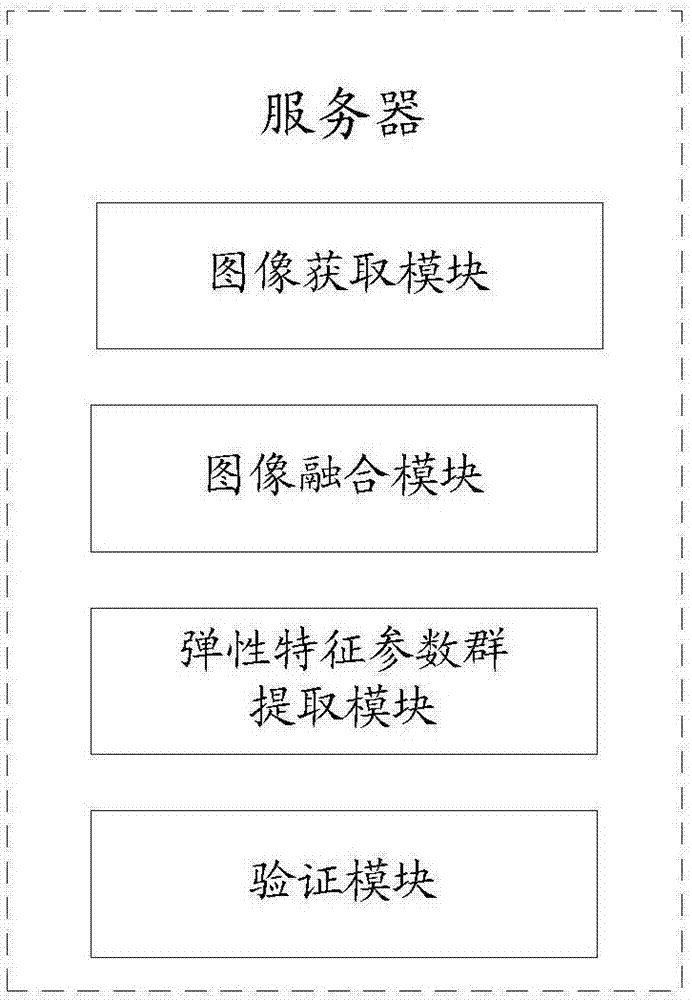
[0041] An embodiment of the tumor image analysis and processing device of the present application is used to combine the hardness information of the ultrasound imaging with the blood oxygen information provided by the photoacoustic imaging to analyze the tumor image.

[0042] Such as figure 2 As shown, the tumor image analysis and processing device of the present application may include an image acquisition module, an image fusion module, an elastic feature parameter group extraction module, and a verification module.

[0043] An image acquisition module, configured to acquire an elastic image of the detected part through ultrasonic shear wave elastography;

[0044] The image fusion module is used to obtain the B-ultrasound image of the detected part, extract the edge contour information of the tumor in the B-ultrasound image, and fuse it to the elastic image to segment the lesion area;

[0045] The elastic feature parameter group extraction module is used to quantify and ex...


PUM


Abstract

The invention provides an analyzing and processing method and device for a tumor image. The method comprises the step of combining hardness information of ultrasonic imaging and blood oxygen information provided by photoacoustic imaging for analysis of the tumor image. In the specific embodiment of the invention, the hardness information of ultrasonic imaging and the blood oxygen information provided by photoacoustic imaging are combined for analysis of the tumor image, critical function and molecular imaging information provided by the photoacoustic technique can be importantly complementarywith tissue morphology and structure information provided by the ultrasonic technique, and overall observation of tumor structure and function is realized through combination of the photoacoustic technique and the ultrasonic technique.

Description

technical field [0001] The present application relates to image processing, in particular to a tumor image analysis and processing method and device. Background technique [0002] With the emergence of quantitative detection technology, elastography has entered a new stage of development. The quantitative detection technology of tissue elasticity mainly includes Fibroscan technology, Acoustic Radiation Force Impulse (ARFI) and shear wave elastography (Shear Wave elastography). Elastic Imaging, SWEI), because Fibroscan technology is a one-dimensional elastic detection technology, there are many studies on the degree of liver fibrosis, and many scholars have used ARFI technology and SWE technology to conduct extensive and in-depth research on the elastic quantitative parameters of tumors in various organs. Analysis, the research results show the good clinical value of elastic quantitative analysis in tumor diagnosis. ARFI technology indirectly judges tissue elasticity by detec...

Claims


Application Information

Patent Timeline
Patent Type & Authority Applications(China)
IPC IPC(8): G06T7/00G06T7/11
CPCG06T7/0012G06T2207/10132G06T2207/30096G06T7/11
Inventor 罗慧徐金锋李剑
Owner SHENZHEN PEOPLES HOSPITAL
Why Eureka
Try Eureka