Cord blood regulative T-cell in-vitro amplification method
A regulatory and cell body technology, applied in the direction of blood/immune system cells, animal cells, vertebrate cells, etc., can solve the problems of high cost and biological safety.
- Summary
- Abstract
- Description
- Claims
- Application Information
AI Technical Summary
Problems solved by technology
Method used
Image

Examples
Embodiment 1
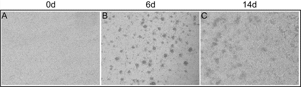
[0026] Example 1 In vitro expansion and culture of Tregs cells derived from fresh umbilical cord blood
[0027] Coat the culture dish with 5 μg / ml purified anti-CD3 antibody solution: dilute the purified anti-CD3 antibody to 5 μg / ml with PBS buffer, add 6 ml of 5 μg / ml anti-anti-CD3 antibody solution to the 100mm culture dish, Place in a 37°C incubator for 4 hours, and discard the antibody. Rinse the Petri dish once with PBS buffer, and air dry the bottom of the Petri dish in the operating table.
[0028] Umbilical cord blood was taken from the umbilical cord of full-term healthy newborns, anticoagulated with sodium citrate, and separated within 24 hours after collection.
[0029] The umbilical cord blood was diluted with PBS at a ratio of 1:1, slowly added to an equal volume of Ficoll lymphocyte separation medium, centrifuged at 2000 rpm for 20 min, at a speed up of 1, and down in a speed of 1. Collect the mononuclear cell buffy coat into a new 50ml centrifuge tube, centrif...
Embodiment example 2
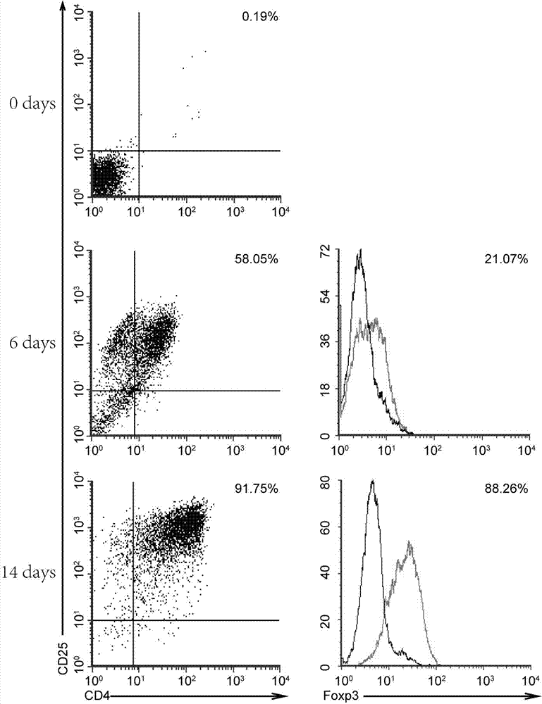
[0034] Example 2 Flow cytometric detection of cell surface antigens obtained from in vitro amplification of fresh cord blood
[0035] The amplified cells were harvested, resuspended in PBS buffer, and counted. Take some cells through a 100-mesh cell sieve, centrifuge, collect the cell pellet, resuspend the pellet with PBS buffer, and adjust it to 1×10 6 cells / ml. Add 200 μl of cell suspension to each tube, and add 5 μL of flow cytometry antibody. Use FITC-labeled anti-CD4 antibody and PE-labeled anti-CD25 antibody to label cell surface antigens at room temperature for 30 minutes. Cells are treated with permeabilization solution and fixative solution for 1 hour. Afterwards, the intracellular antigen was labeled with Alexa488-labeled anti-Foxp3 antibody for 30 min. The result is as figure 2 showed that CD4 + CD25 + The proportion of T cells was only 0.19% compared to ( figure 2 , 0day), CD4 in cells obtained after expansion + CD25 + The proportion of T cells increased ...
Embodiment 3
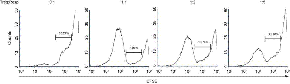
[0036] Example 3 Amplified and Obtained Cells Inhibition Effector Cell Proliferation Function Detection
[0037] Mononuclear cells in adult peripheral blood were separated by Ficoll lymphocyte separation medium density gradient centrifugation, and CD4 obtained by immunomagnetic bead separation and purification + CD25 - Cells were used as effector T cells, after staining with CFSE (5 μM), 1×10 5 Cells / well were seeded in anti-CD3 antibody-coated 96-well plates, according to the ratio of Tregs: effector T cells = 0:1, 1:1, 1:2, 1:5, the expanded cells were added to the 96-well plates Fresh umbilical cord blood Tregs cells were added to each well with Gibco RPMI 1640 + 10% FBS + 2000IU / ml IL-2 + 100ng / ml anti-CD28 culture solution, and cultured at 37°C, 5% and saturated humidity for 4 days, 4 The cells in the 96-well plate were collected one day later, and the results were analyzed by flow cytometry. Such as image 3 As shown, Tregs cells obtained by in vitro expansion can ef...
PUM
 Abstract
Description
Claims
Application Information
 - R&D
- Intellectual Property
- Life Sciences
- Materials
- Tech Scout
- Unparalleled Data Quality
- Higher Quality Content
- 60% Fewer Hallucinations
Browse by: Latest US Patents, China's latest patents, Technical Efficacy Thesaurus, Application Domain, Technology Topic, Popular Technical Reports.
© 2025 PatSnap. All rights reserved.Legal|Privacy policy|Modern Slavery Act Transparency Statement|Sitemap|About US| Contact US: help@patsnap.com