Novel cervical exfoliated cell separation method

A technique for exfoliated cells and cervix, applied in the field of cervical exfoliated cell separation, can solve the problems affecting the detection rate of abnormal cells, small cells, large nuclei, etc., and achieve the effect of high practical application value, far-reaching clinical value and broad market prospect.

Inactive Publication Date: 2010-12-15
ANHUI PROVINCIAL HOSPITAL
View PDF0 Cites 4 Cited by
  • Summary
  • Abstract
  • Description
  • Claims
  • Application Information

AI Technical Summary

Problems solved by technology

[0003] At present, the main methods for screening cervical cancer include cervical smear exfoliation cytology examination, cervical epithelial liquid-based cytology examination, etc., but there are certain limitations. Precancerous lesions are symptoms of a gradual transition from benign to malignant, including: 1. The number of keratinocytes in cervical smears continues to increase, and the cells in the smears are continuously affected by estrogen, and the degree of keratinization is equivalent to the cycle The highest level before mid-ovulation; 2. Atypical or precocious keratinocytes; 3. Huge cells, or small cells; 4. Abnormally large nuclei, active and immature, densely stained nuclear chromatin, often

Method used


Image

  • Novel cervical exfoliated cell separation method
  • Novel cervical exfoliated cell separation method
  • Novel cervical exfoliated cell separation method

Examples


Embodiment Construction

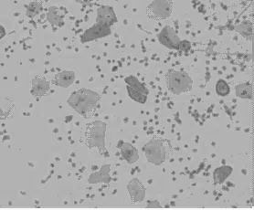
[0027] The method for separating cervical exfoliated cells in the present embodiment operates as follows:

[0028] 1. Use a special brush for cervical exfoliated cells, gently press the brush head on the cervix, and rotate 2-3 times in the same direction to brush the cervical exfoliated cells, and place the brush in 5ml of PBS phosphoric acid with a concentration of 10% by volume Buffer, vortex and mix to obtain cell suspension;

[0029] 2. Add the cell suspension obtained in step 1 into 3ml of mucus separation solution along the tube wall; after centrifugation and discarding the supernatant, add 3ml of PBS phosphate buffer solution with a volume percentage concentration of 10% to the centrifuged sediment. Resuspend the precipitate to obtain a resuspended cell suspension;

[0030] 3. Add the resuspended cell suspension obtained in step 2 into 3ml of neutrophil separation medium Ficoll along the tube wall, centrifuge, discard the supernatant, and the precipitate after centrifu...


PUM


Abstract

The invention discloses a novel cervical exfoliated cell separation method, which is characterized by: placing cervical exfoliated cells in phosphate buffer solution (PBS) and uniformly mixing by whirling to obtain cell suspension; adding the cell suspension into mucilage separating solution; centrifuging, removing supernate, adding the phosphate buffer solution (PBS) again and re-suspending sediment to obtain re-suspended cell suspension; and adding the re-suspended cell suspension into neutral granular cell separating solution Ficoll, centrifuging, removing supernate and obtaining the settlement which is pure cells after centrifugation. The method of the invention greatly purify clinically extracted cervical exfoliated cell specimens, adopts simple and convenient operation steps, is suitable for large-scale cell separation with stable and reliable effect, can obtain high-purity target cells, has high actual use value and a promising market prospect.

Description

technical field [0001] The invention relates to a method for separating cervical exfoliated cells. Background technique [0002] Cervical cancer is one of the common malignant tumors in gynecology. Its incidence rate is second only to breast cancer, ranking second in female malignant tumors, and its mortality rate ranks first in gynecological malignant tumors. In recent years, cervical cancer patients have been increasing at an annual rate of 2% to 4%. At the same time, compared with developed countries, the incidence of cervical cancer in developing countries is 6 times that of developed countries due to imperfect cervical screening. , and 80% of the patients were diagnosed with invasive cancer, and cervical cancer has a long period of reversible precancerous lesions, so early detection of precancerous lesions is the key to prevent and treat cervical cancer. [0003] At present, the main methods for screening cervical cancer include cervical smear exfoliation cytology exam...

Claims


Application Information

Patent Timeline
IPC IPC(8): C12N5/071
Inventor 凌斌周颖李敏许乾乾冯定庆朱园园沈国栋李彩荣卫莹雷蕾王青元朱靖姚凤球
Owner ANHUI PROVINCIAL HOSPITAL
Why Eureka
Try Eureka