Method for automatically detecting coronary artery calcified plaque of human heart

A technology for automatic detection of calcified plaque, applied in the field of medical image processing, can solve the problem of taking a long time, and achieve the effect of simple parameters, guaranteed accuracy, and comprehensive extraction results

Active Publication Date: 2018-06-15
数坤(上海)医疗科技有限公司
View PDF5 Cites 72 Cited by
  • Summary
  • Abstract
  • Description
  • Claims
  • Application Information

AI Technical Summary

Problems solved by technology

However, the early detection and quantification of plaque requires very experienced doctors to spend a long time on manual plaque segmentation and analysis. Therefore, it is necessary to propose an automatic and rapid detection method for cardiac coronary calcified plaque to improve the work efficiency of doctors.
[00...

Method used


Image

  • Method for automatically detecting coronary artery calcified plaque of human heart
  • Method for automatically detecting coronary artery calcified plaque of human heart
  • Method for automatically detecting coronary artery calcified plaque of human heart

Examples


Embodiment

[0046] refer to figure 1 As shown, a method for automatically detecting calcified plaques in human heart coronary arteries comprises the following steps:

[0047] S1. Use the deep learning neural network to segment the original image of the coronary CTA sequence to obtain the extracted image of the coronary artery of the human heart;

[0048] S2. Process the extracted image of the coronary arteries of the human heart to generate a straightened image of each branch vessel;

[0049] S3. Carry out blood vessel segmentation on each straightened picture, and obtain the straightened blood vessel map of each branch blood vessel;

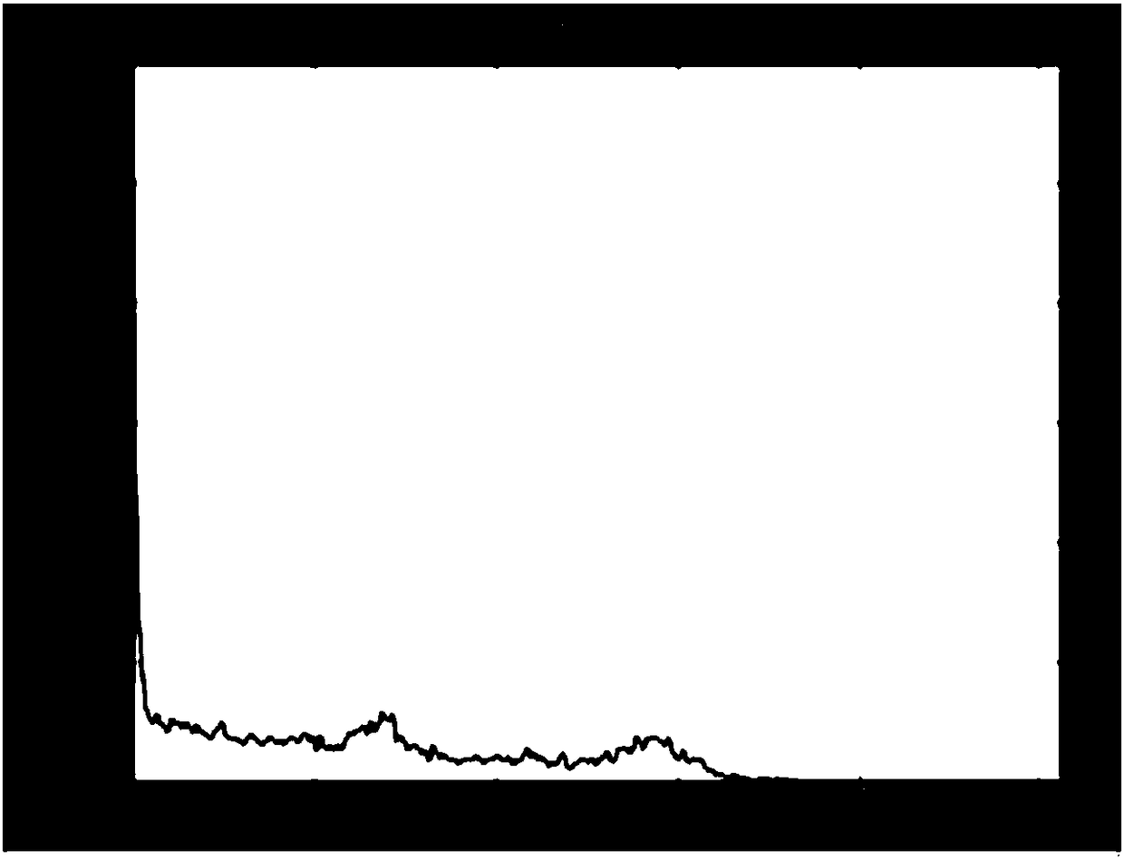
[0050] S4. Adjust the window frame and window level, and calculate the pixel value of the entire image for each straightened blood vessel map. If there are pixels with a pixel value greater than 220, it is determined that there is a calcified plaque, which is screened out from the straightened blood vessel map. Diagram of a straightened blood vessel with ...


PUM


Abstract

The invention discloses a method for automatically detecting a coronary artery calcified plaque of a human heart. The method comprises the following steps of S1.adopting a deep learning neural networkto segment an original graph of a coronary artery CTA sequence in order obtain a coronary artery extraction graph of the human heart; S2.processing the coronary artery extraction graph of the human heart to generate a straightening picture of each branch vessel; S3.carrying out blood vessel segmentation on each straightening picture to obtain a straightening blood vessel graph of each branch vessel; S4.adjusting a window width and a window level, calculating a pixel value of the whole picture of each straightening blood vessel graph, if a pixel point whose pixel value is greater than 220 exists, determining that a calcified plaque exists, and screening out the straightening blood vessel graph with the calcified plaque; S5.converting the straightening blood vessel graph with the calcifiedplaque into a grey scale graph, filling the pixel point whose gray value is larger than 220 with the color, and obtaining a calcified plaque extraction result; and S6.calculating a rate of stenosis ofthe blood vessel and obtaining a quantization value. The method is effective for detection of most calcified plaques, automatic detection can be realized, and the efficiency is greatly improved.

Description

technical field [0001] The invention relates to the field of medical image processing, in particular to a method for automatically detecting calcified plaques in human heart coronary arteries. Background technique [0002] Finding a safe and reliable method for detecting coronary artery disease is the main goal of clinical development in the future, so it is of great clinical value and practical significance to be able to accurately extract plaque from CTA image sequences to judge coronary artery disease. In the past ten years, the proportion of deaths caused by coronary artery disease has been increasing year by year. Therefore, it is necessary to accurately extract and quantify arterial vessels, especially the early detection and quantitative analysis of plaque. However, the early detection and quantification of plaque requires very experienced doctors to spend a long time on manual plaque segmentation and analysis. Therefore, it is necessary to propose an automatic and ra...

Claims


Application Information

Patent Timeline
IPC IPC(8): G06T7/00G06T7/10
CPCG06T7/001G06T7/10G06T2207/10016G06T2207/20081G06T2207/20084G06T2207/30048G06T2207/30101
Inventor 穆红章马春娥安宝磊
Owner 数坤(上海)医疗科技有限公司
Why Eureka
Try Eureka