

Prostatic cancer diagnosis developer and preparation method thereof
A technology for diagnosing imaging and prostate cancer, applied in the field of nuclear medicine, can solve the problems of false positives, low specificity, low sensitivity, etc., and achieve the effects of high uptake, simple labeling method, high sensitivity and specificity
- Summary
- Abstract
- Description
- Claims
- Application Information
AI Technical Summary
Problems solved by technology
Method used
Image

Examples
Embodiment 1
[0021] Using a proton cyclotron 18 O(p,n) 18 The F reaction produces an activity of 100mCi 18 f - ; the resulting 18 f - Transfer to the anion exchange column QMA through pipeline (QMA is READI-CLING TM type column, the column body is hollowed out, and about 1 / 5Plus C18 column filler is refilled) to capture, after the capture is completed, use N 2 Blow dry the QMA, then rinse with 0.3ml PH=4.2 acetic acid-sodium acetate buffer solution, elute to get 18 f - The buffer solution, its activity is 90mCi;
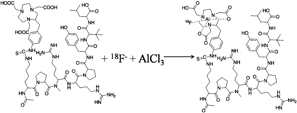
[0022] to 0.3ml 18 f - Add 300ug NOTA-modified neurotensin, 0.5ml acetonitrile and 0.03ml 0.4M AlCl to the buffer solution 3 solution and reacted at 80°C for 10min, then added 5ml of deionized water to quench the reaction, and then passed the reaction solution over Al 2 o 3 Column, obtain filtrate;
[0023] Pass the filtrate through C 18 Plus column was enriched, and then rinsed with 2mL ethanol and 10mL normal saline in sequence, and the rinse solution was passed t...
Embodiment 2
[0039] Using a proton cyclotron 18 O(p,n) 18 The F reaction produces an activity of 100mCi 18 f - ,; then the generated 18 f - Transfer to the anion exchange column QMA through pipeline (QMA is READI-CLING TM type column, the column body is hollowed out, and about 1 / 5Plus C18 column filler is refilled) to capture, after the capture is completed, use N 2 Blow dry the QMA, then rinse with 0.3ml PH=4.2 acetic acid-sodium acetate buffer solution, elute to get 18 f - The buffer solution, its activity is 80mCi;
[0040] to 0.3ml 18 f - Add 300ug NOTA-modified neurotensin, 0.5ml acetonitrile and 0.03ml 0.4M AlCl to the buffer solution 3 solution and reacted at 80°C for 10min, then added 5ml of deionized water to quench the reaction, and then passed the reaction solution over Al 2 o 3 Column, obtain filtrate;
[0041] Pass the filtrate through C 18 Plus column was enriched, and then rinsed with 2mL ethanol and 10mL normal saline in sequence, and the rinse solution was pa...
Embodiment 3
[0043] Using a proton cyclotron 18 O(p,n) 18 The activity of F reaction is 110mCi 18 f - ,; then the generated 18 f - Transfer to the anion exchange column QMA through pipeline (QMA is READI-CLING TM type column, the column body is hollowed out, and about 1 / 5Plus C18 column filler is refilled) to capture, after the capture is completed, use N 2 Blow dry the QMA, then rinse with 0.3ml PH=4.2 acetic acid-sodium acetate buffer solution, elute to get 18 f - The buffer solution, its activity is 84mCi;
[0044] to 0.3ml 18 f - Add 300ug NOTA-modified neurotensin, 0.5ml acetonitrile and 0.03ml 0.4M AlCl to the sodium acetate solution 3 solution and reacted at 80°C for 10min, then added 5ml of deionized water to quench the reaction, and then passed the reaction solution over Al 2 o 3 Column, obtain filtrate;
[0045] Pass the filtrate through C 18 Plus column was enriched, and then rinsed with 2mL ethanol and 10mL normal saline in sequence, and the rinse solution was pas...
PUM
 Abstract
Description
Claims
Application Information