
Eyeground image processing method and device, storage medium, and processor

A fundus image and processing method technology, which is applied in the medical field, can solve the problem that the fundus image cannot be effectively registered, and achieve the effect of effective registration

Inactive Publication Date: 2018-06-22
海纳医信(北京)软件科技有限责任公司
View PDF3 Cites 9 Cited by
  • Summary
  • Abstract
  • Description
  • Claims
  • Application Information

AI Technical Summary

Problems solved by technology

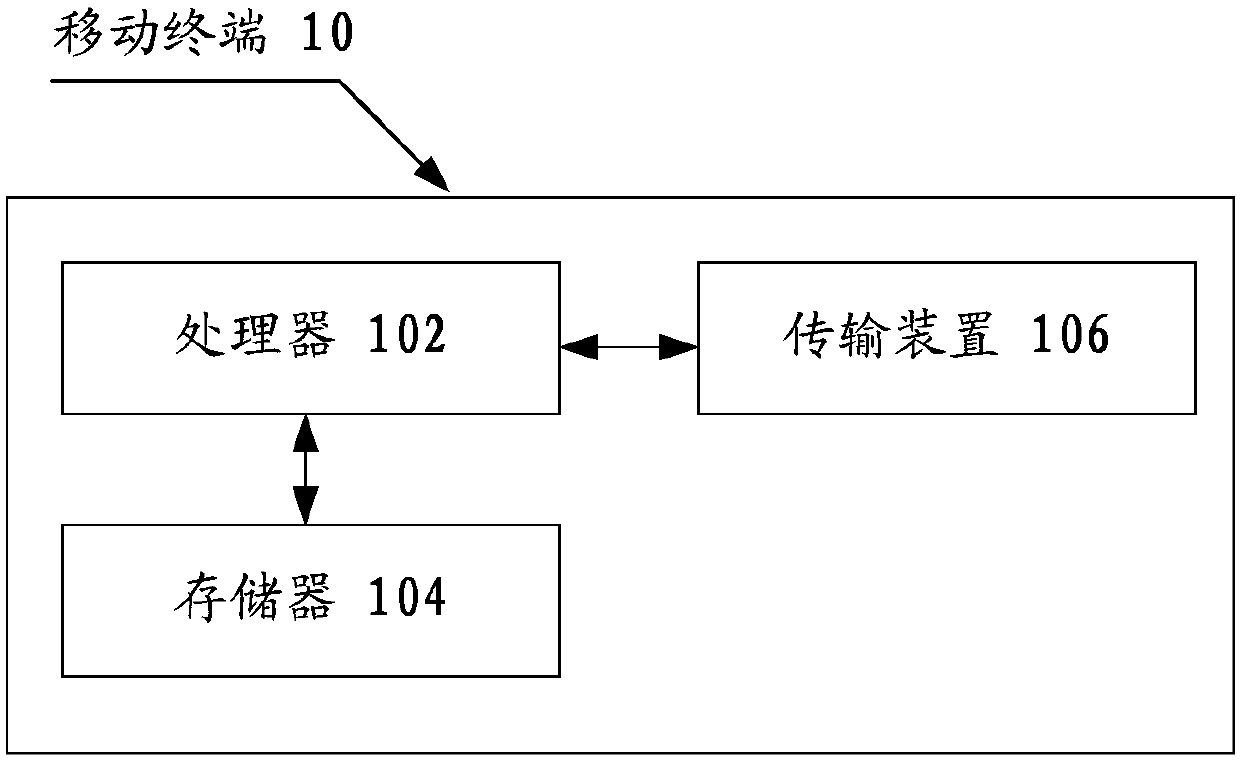
[0004] Embodiments of the present invention provide a fundus image processing method and device, a storage medium, and a processor to at least solve the problem in the related art that effective registration of fundus images based on blood vessel features cannot be performed.

Method used


Image

  • Eyeground image processing method and device, storage medium, and processor
  • Eyeground image processing method and device, storage medium, and processor
  • Eyeground image processing method and device, storage medium, and processor

Examples


specific Embodiment 1

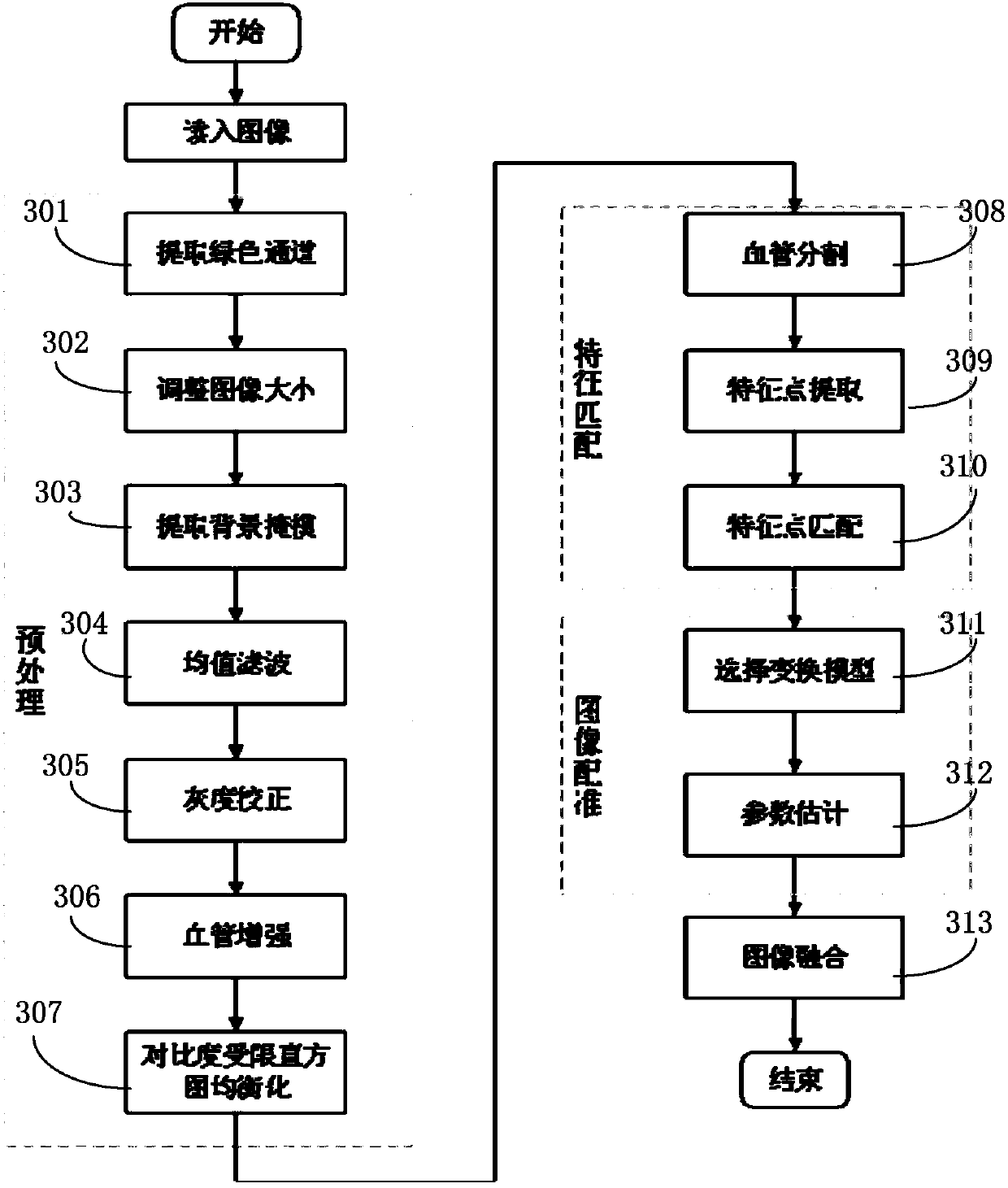
[0043] The blood vessel structure is a relatively stable and widely covered physiological structure in the fundus image, and the bifurcation point of the blood vessel is invariant to the gray scale and illumination changes, so this embodiment proposes a fundus image registration method based on blood vessel features. Firstly, the fundus image is preprocessed, and the fundus image is segmented by Hessian matrix and morphological method; secondly, on the basis of blood vessel segmentation, the relevant information (such as: coordinates, entropy, etc.) value, geodesic distance and Euclidean distance, radon transformation result) as the feature point set; then, find out the initial matching point that satisfies the mapping condition, and purify the initial matching point to determine the best matching point; finally, use affine Transformation is used to register and fuse images.

[0044] image 3 is a flow chart in the embodiment of the present invention, such as image 3 As sho...


PUM


Abstract

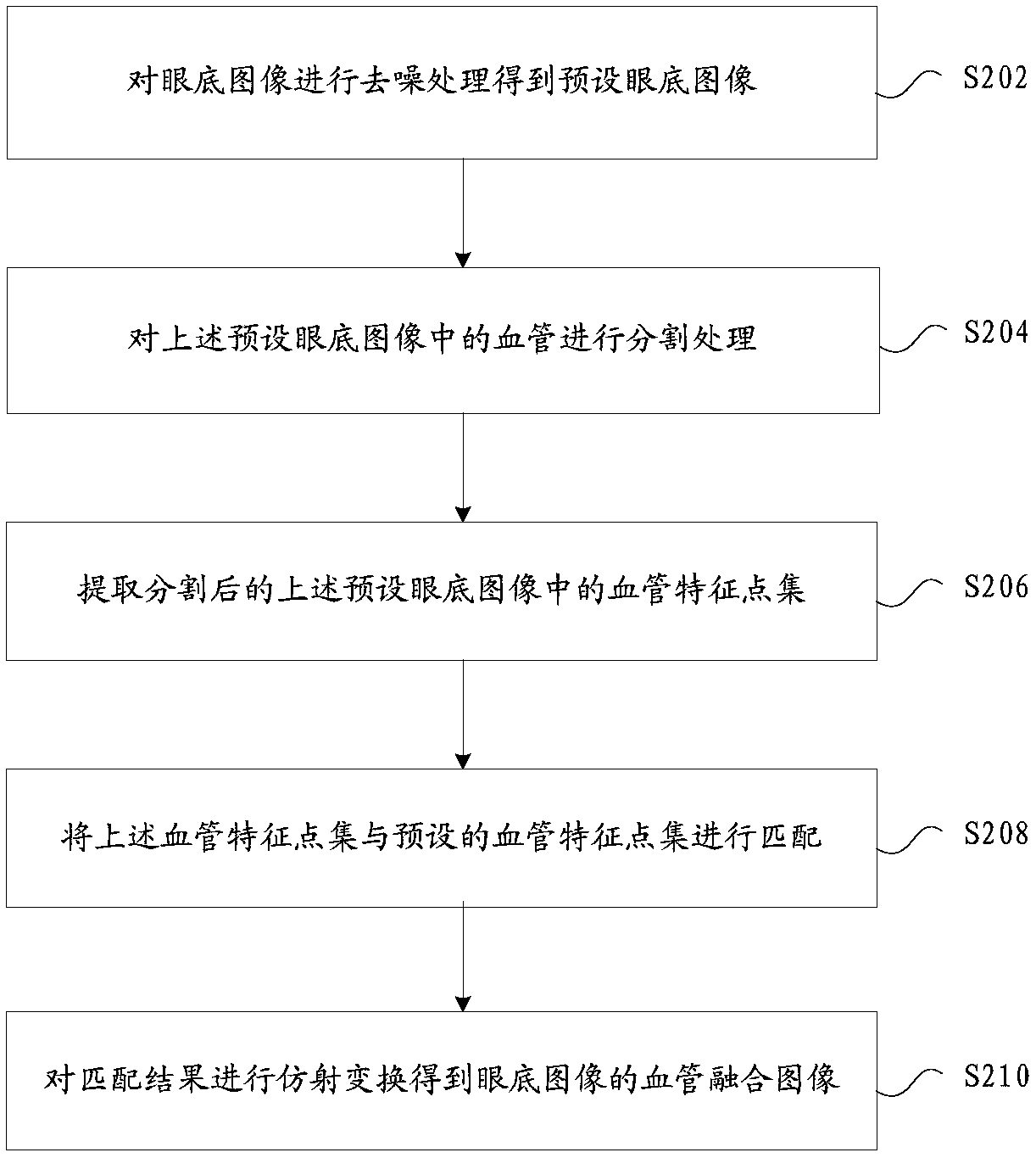
The invention provides an eyeground image processing method and device, a storage medium, and a processor. The method comprises the steps: carrying out the denoising of an eyeground image, and obtaining a preset eyeground image; carrying out the segmentation of a blood vessel in the preset eyeground image; extracting a blood vessel feature set in the preset eyeground image after segmentation; carrying out the matching of the blood vessel feature set with a preset eyeground image set; carrying out the affine transformation of a matching result, and obtaining a blood vessel fusion image of the eyeground image. According to the invention, the method can solve a problem that an eyeground image based on the blood vessel features cannot be effectively registered, and achieves the effective registration of the eyeground image according to the blood vessel features.

Description

technical field [0001] The present invention relates to the medical field, in particular to a fundus image processing method and device, a storage medium, and a processor. Background technique [0002] The vascular structure is a relatively stable and widely covered physiological structure in fundus images, and the bifurcation points of blood vessels are invariant to changes in grayscale and light. In the actual diagnosis and treatment process, doctors may have inaccurate diagnoses due to experience and other reasons. In the prior art, effective registration cannot be performed on fundus images based on blood vessel features. [0003] For the above-mentioned problems, no effective solution has been proposed in the related art. Contents of the invention [0004] Embodiments of the present invention provide a fundus image processing method and device, a storage medium, and a processor, so as to at least solve the problem in the related art that effective registration of fun...

Claims


Application Information

Patent Timeline
IPC IPC(8): G06T7/33G06T7/11G06T5/00G06T5/40
CPCG06T5/40G06T7/11G06T7/337G06T2207/30041G06T2207/20221G06T5/70
Inventor 崔彤哲周永新陈国桢孙毅刘萍
Owner 海纳医信(北京)软件科技有限责任公司
Learn More