Fundus image blood vessel feature extraction method, device and system and storage medium

A fundus image and extraction method technology, applied in image enhancement, image analysis, image data processing and other directions, can solve problems such as unrealized management and utilization

Active Publication Date: 2020-03-10
FUZHOU YIYING HEALTH TECH CO LTD
View PDF6 Cites 3 Cited by
  • Summary
  • Abstract
  • Description
  • Claims
  • Application Information

AI Technical Summary

Problems solved by technology

[0003] At present, many countries in the world, including my country, have carried out the construction of community-based fundus screening syst

Method used


Image

  • Fundus image blood vessel feature extraction method, device and system and storage medium
  • Fundus image blood vessel feature extraction method, device and system and storage medium
  • Fundus image blood vessel feature extraction method, device and system and storage medium

Examples


Embodiment Construction

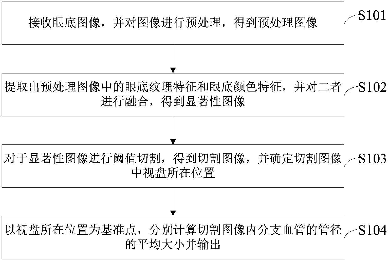
[0054] In order to explain in detail the technical content, structural features, achieved goals and effects of the technical solution, the following will be described in detail in conjunction with specific embodiments and accompanying drawings.

[0055] see figure 1 , is a schematic diagram of a method for extracting blood vessel features of a fundus image according to an embodiment of the present invention. The method comprises the steps of:

[0056]First enter step S101 to receive the fundus image, and preprocess the image to obtain the preprocessed image. As the name implies, a fundus image is an image that contains fundus information. The fundus is the tissue at the back of the eyeball, that is, the inner membrane of the eyeball—including the retina, optic disc, macula, and central retinal artery and vein. Fundus images can be collected with a color fundus camera.

[0057] In the process of practical application, considering that the collected fundus images are often di...


PUM


Abstract

The invention provides a fundus image blood vessel feature extraction method, device and system and a storage medium, and the method comprises the following steps: receiving a fundus image, and carrying out the preprocessing of the image, and obtaining a preprocessed image; extracting fundus texture features and fundus color features in the preprocessed image, and fusing the fundus texture features and the fundus color features to obtain a saliency image; carrying out threshold value cutting on the saliency image to obtain a cut image, and determining the position of a visual disk in the cut image; taking the position of the optic disc as a reference point, respectively calculating the average size of the pipe diameters of the branch blood vessels in the cutting image and outputting. According to the invention, the fundus image can be acquired, and the change of retinal vessel characteristics is automatically analyzed, so that the small vessel lesion condition is discovered in time, auxiliary diagnosis information is provided for doctors, and personalized health services of hypertension and other chronic diseases under precision medicine are realized.

Description

technical field [0001] The invention relates to the fields of fundus image analysis and lesion feature extraction, in particular to a fundus image blood vessel feature extraction method, device, system and storage medium. Background technique [0002] Eyes are the windows of health, and the only part of the human body where blood vessels can be directly seen without surgery. Medical evidence shows that the retina and the brain's circulatory system share similar anatomical, physiological, and embryonic developmental characteristics. Therefore, through the fundus blood vessels, we can understand the degree of disease in the whole body, especially the brain arteries and small and medium-sized arteries throughout the body. The changes in the fundus blood vessels are also the main basis for risk stratification of hypertension. The status and changes of the retina, retinal blood vessels, and optic nerve head reflect to a certain extent The dynamic situation and health status of t...

Claims


Application Information

Patent Timeline
IPC IPC(8): G06K9/46G06K9/62G06T7/11G06T7/41G06T7/62G06T7/90
CPCG06T7/11G06T7/41G06T7/90G06T7/62G06T2207/30041G06T2207/30101G06V10/462G06V10/56G06F18/253
Inventor 余轮薛岚燕林嘉雯曹新容王丽纳
Owner FUZHOU YIYING HEALTH TECH CO LTD
Why Eureka
Try Eureka