Cardiac anatomic structure-based left and right ventricle level set segmentation method

A technology of level set segmentation and cardiac anatomy, applied in the field of medical imaging, can solve problems such as lack, and achieve the effect of sub-pixel accuracy

Inactive Publication Date: 2018-06-29
UNIV OF ELECTRONICS SCI & TECH OF CHINA
View PDF5 Cites 10 Cited by
  • Summary
  • Abstract
  • Description
  • Claims
  • Application Information

AI Technical Summary

Problems solved by technology

However, the current segmentation algorithm for cardiac images is mainly for the segmentation of a single cardiac structure, and there is a lack of alg

Method used


Image

  • Cardiac anatomic structure-based left and right ventricle level set segmentation method
  • Cardiac anatomic structure-based left and right ventricle level set segmentation method
  • Cardiac anatomic structure-based left and right ventricle level set segmentation method

Examples


Embodiment Construction

[0039] Describe below in conjunction with accompanying drawing:

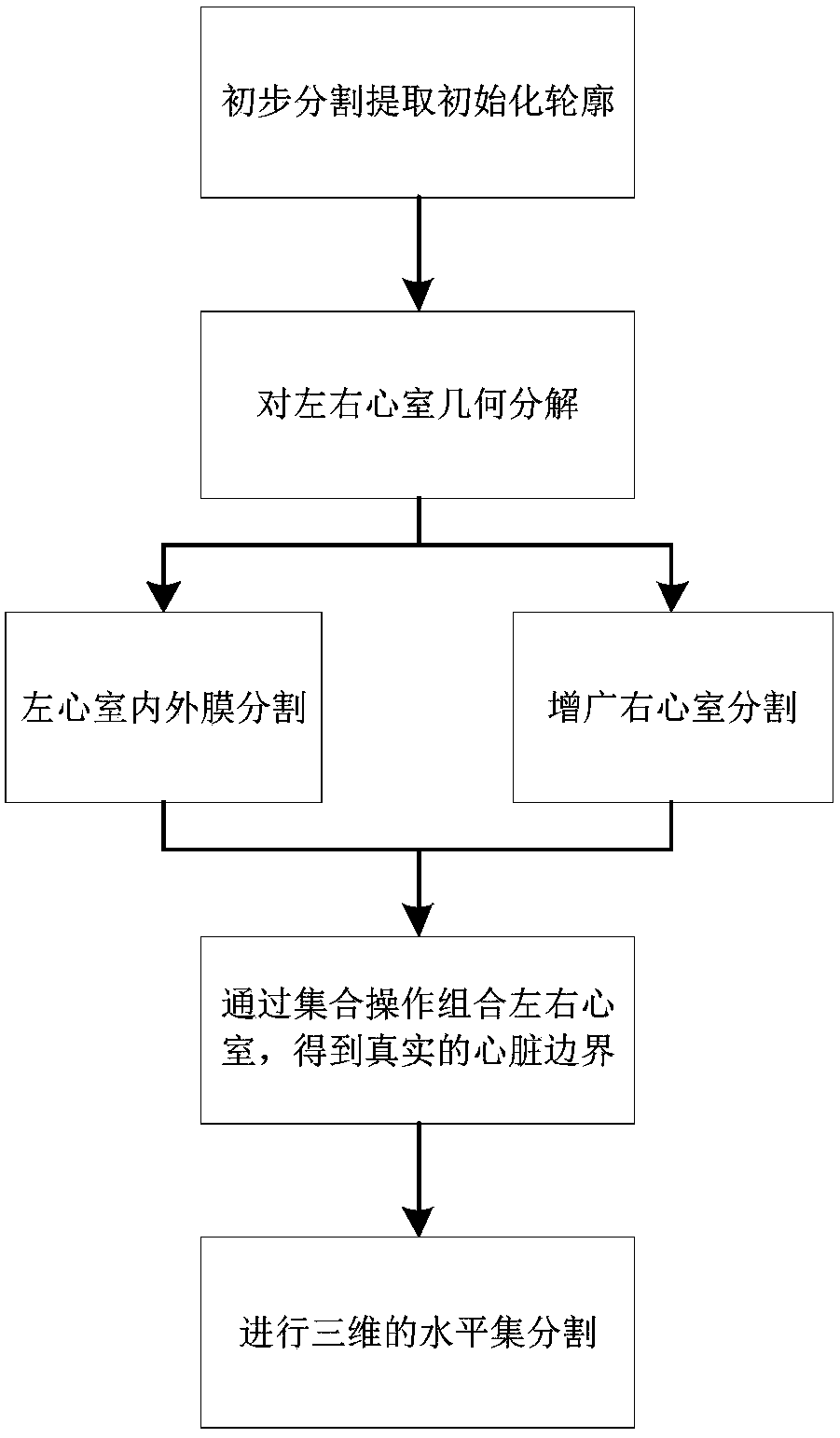
[0040] A level set segmentation method of left and right ventricles based on cardiac anatomy:

[0041] S1. Preliminarily segment the cardiac MRI (Cardiovascular Magnetic Resonance, CMR) to obtain a binary image including the heart and other tissues around the heart. Manually determine the position of the center point of the left and right ventricles in an interactive manner, and extract the Initialize the outline of the binary image of the left and right ventricles; firstly, perform step S1 to perform preliminary segmentation on the two-dimensional magnetic resonance image, and obtain a binary image including the heart and other tissues around the heart. By interactively determining the positions of the center points of the left and right ventricles, the binary image initialization contours of the left and right ventricles can be extracted according to the positions of the center points.

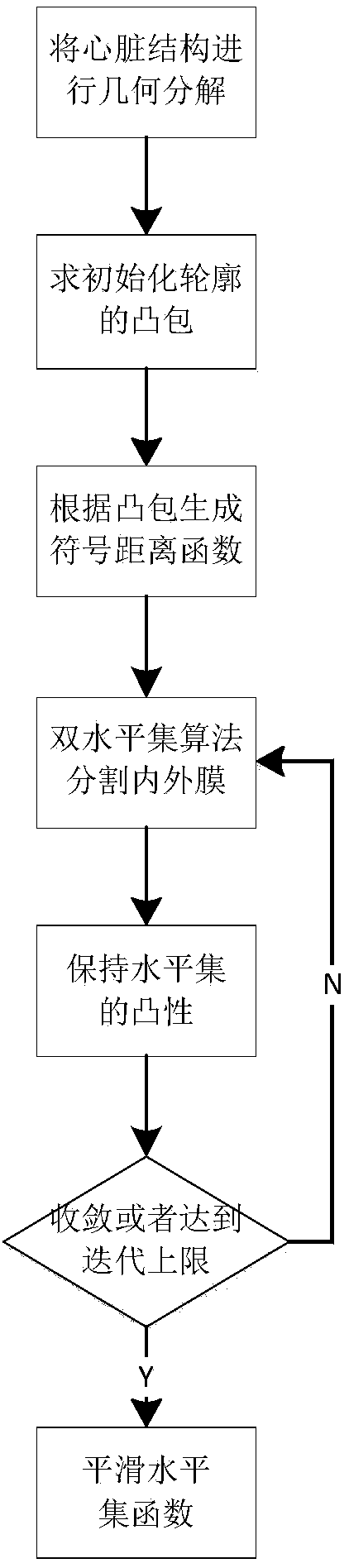
[0042] Among them, th...


PUM


Abstract

The invention belongs to the technical field of medical imaging, and particularly relates to a cardiac magnetic resonance image segmentation method. A cardiac anatomic structure-based left and right ventricle level set segmentation method specifically comprises the following steps: performing geometry decomposition to a cardiac structure; resolving a convex hull of an initialized contour; producing a signaled distance function; and generating intima and extima through a dual-level set algorithm, and the like. By using the method, the simultaneous segmentation of the ventricular intima and extima and cardiac muscle can be realized, and the segmentation results can meet the physiological structure characteristics of a heart as far as possible.

Description

technical field [0001] The invention relates to the technical field of medical imaging, in particular to a method for segmenting cardiac magnetic resonance images. Background technique [0002] In the diagnosis of heart disease, medical imaging technology has become an important means of clinical examination. Among them, magnetic resonance imaging is considered the "gold standard" for noninvasive assessment of cardiac structure and function. In the analysis and processing of magnetic resonance images, accurate segmentation of cardiac anatomical structure is the basis and premise for functional analysis and disease diagnosis of the heart. However, due to the image quality problems caused by the gray scale inhomogeneity of magnetic resonance imaging, the complexity of the physiological structure of the heart and the blurring of the border caused by the heart movement, it is difficult for the classical image segmentation method to accurately segment the cardiac magnetic resona...

Claims


Application Information

Patent Timeline
IPC IPC(8): G06T7/12G06T7/13G06T7/187G06T7/64G06T7/00
CPCG06T7/0012G06T2207/10088G06T2207/30048G06T7/12G06T7/13G06T7/187G06T7/64
Inventor 李纯明
Owner UNIV OF ELECTRONICS SCI & TECH OF CHINA
Why Eureka
Try Eureka