
Method for automatically removing muscle artifacts in single-channel EEG signal

An EEG signal, single-channel technology, used in medical science, sensors, diagnostic recording/measurement, etc., can solve problems such as data loss, loss of EEG signals, and not suitable for real-time processing, and achieve the best real-time performance and running time. short effect

Inactive Publication Date: 2018-07-24
SOUTH CHINA UNIV OF TECH
View PDF4 Cites 17 Cited by
  • Summary
  • Abstract
  • Description
  • Claims
  • Application Information

AI Technical Summary

Problems solved by technology

Artifact rejection is to remove the entire EEG period containing artifacts. The main disadvantage of this method is that it will also eliminate important EEG information, resulting in data loss; and artifact correction refers to the use of various methods to remove artifact components while retaining as much EEG information as possible
Artifact correction technology mainly includes: 1) filtering, which removes EMG artifacts in EEG signals by using filters of specific frequency bands; Therefore, while removing myoelectric artifacts, a part of the EEG signal will also be lost.
2) Wavelet transform (wavelet transform, WT), the observed signal is decomposed into a series of basis functions by wavelet transform, and then the coefficients are thresholded to remove myoelectric artifacts, and finally the pure EEG signal is obtained by wavelet reconstruction; but wavelet Transformation methods require extensive experimentation to select appropriate wavelet basis functions and decomposition levels
EMD is a data-driven method for processing non-stationary, nonlinear, and stochastic processes, so it is very suitable for EEG signal analysis and processing, but EMD has high computational complexity and generally takes several minutes, which is not easy. suitable for real-time processing

Method used


Image

  • Method for automatically removing muscle artifacts in single-channel EEG signal
  • Method for automatically removing muscle artifacts in single-channel EEG signal
  • Method for automatically removing muscle artifacts in single-channel EEG signal

Examples


Embodiment Construction

[0021] Specific embodiments of the present invention will be described below in conjunction with the accompanying drawings, so that those skilled in the art can better understand the present invention, but the embodiments of the present invention are not limited thereto. It should be noted that in the following description, when detailed descriptions of known functions and designs may dilute the main content of the present invention, these detailed descriptions will be omitted here.

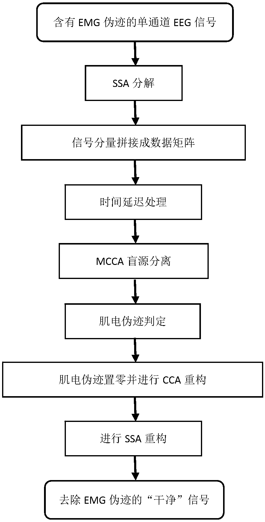
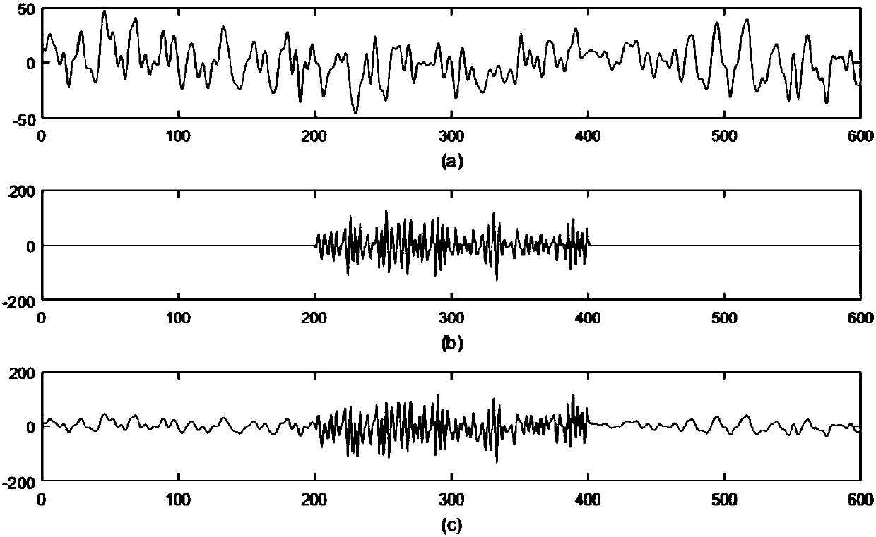
[0022] In this embodiment, the semi-simulated EEG signal is taken as an example, and its flow chart can be found in figure 1 , including the following steps:

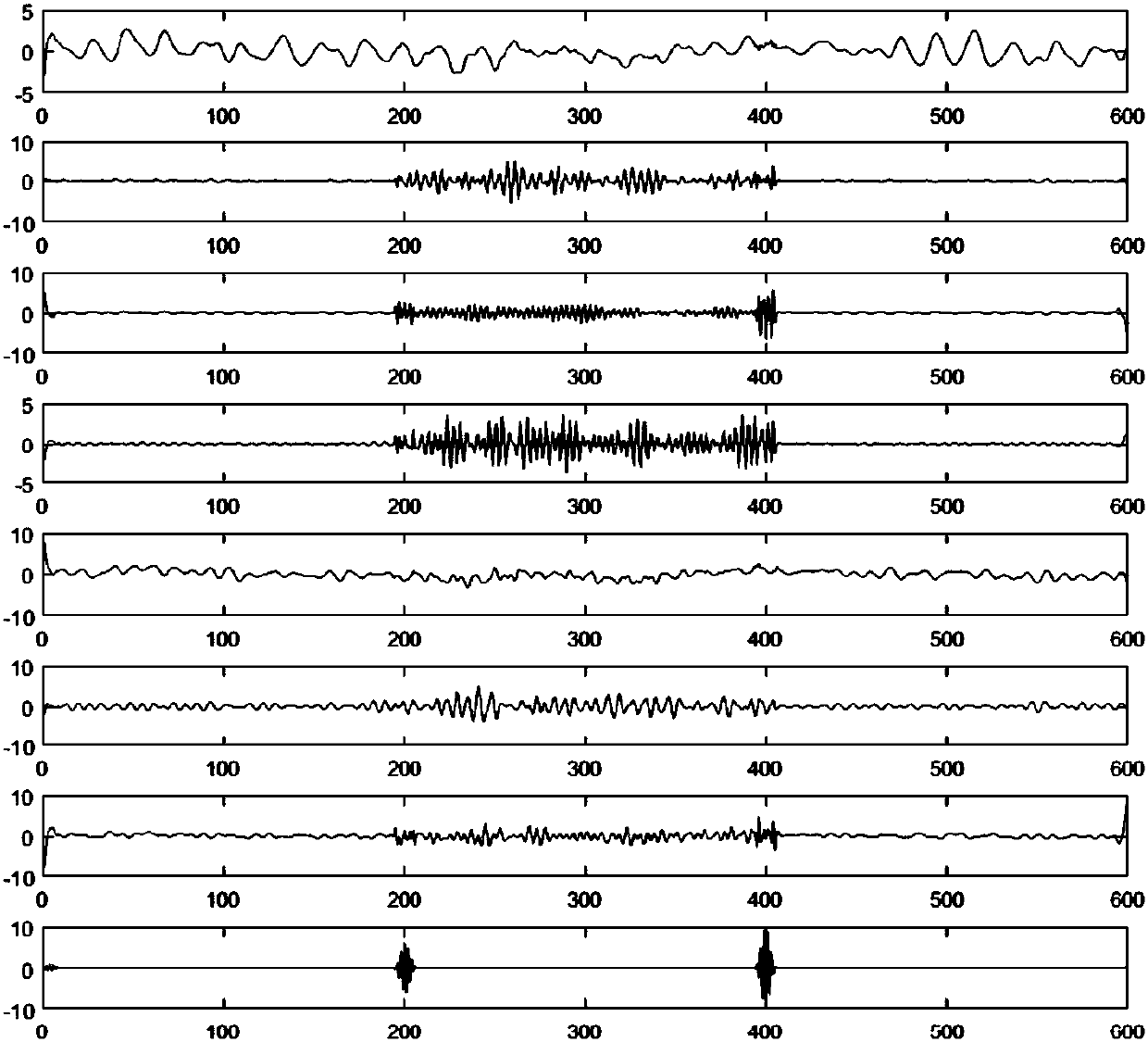
[0023] Step 1: Decompose the EEG signal collected by the single-channel EEG electrode sensor through the Singular Spectrum Analysis (SSA) algorithm to obtain P signal components;

[0024] Specifically: assume that the collected single-channel EEG signal containing myoelectric artifacts is x=[x(1),x(2),...,x(T)], wherein the sequence x(t)(...


PUM


Abstract

The invention discloses a method for automatically removing muscle artifacts in a single-channel EEG signal. The method includes the steps that the EEG signal is subjected to SSA decomposition to obtain P signal components; the P signal components are spliced into a P-dimension data matrix by row; the P-dimension data matrix is subjected to time delay processing to obtain several data matrixes; the several data matrixes are subjected to blind source separation with MCCA to obtain a source estimation matrix S and a hybrid matrix A; sources related to muscle artifacts in the source estimation matrix are recognized; muscle artifacts in the source estimation matrix are removed, the sources recognized to be muscle artifacts are set to be zero to obtain a source estimation matrix S' with muscleartifacts eliminated, and a multi-channel EEG signal X'=A*S' with muscle artifacts removed is obtained through reconstruction; all rows of the multi-channel EEG signal X' are summated, and a single-channel EEG signal x' with muscle artifacts removed can be finally obtained. By means of the method, EEG information is retained as far as possible while the muscle artifacts are removed, and thus the accuracy of EEG signal analysis is improved.

Description

technical field [0001] The invention relates to the technical field of electroencephalogram signal processing, in particular to a method for automatically removing myoelectric artifacts in single-channel electroencephalogram signals. Background technique [0002] Electroencephalogram (Electroencephalograph, EEG) is the reflection of the electrophysiological activity of brain nerve cells on the scalp. Due to its high temporal resolution, non-invasiveness, low cost, and suitability for long-term monitoring, EEG has been widely used to study brain function and pathological brain mechanisms. But while EEG records brain activity, it also records electrical signals produced by other activities outside the brain, and these recorded electrical signals that are not produced by brain activity are called artifacts. Common artifacts include Electrooculogram (EOG), Electromyography (EMG), Electrocardiography (ECG), and noise caused by circuits or external devices. Compared with other t...

Claims


Application Information

Patent Timeline
Patent Type & Authority Applications(China)
IPC IPC(8): A61B5/0476A61B5/00
CPCA61B5/7203A61B5/7235A61B5/316A61B5/369
Inventor 陈灿邢晓芬徐向民舒琳
Owner SOUTH CHINA UNIV OF TECH
Learn More