Method and Apparatus Using Electric Field for Improved Biological Assays

Active Publication Date: 2009-02-05
THE BOARD OF TRUSTEES OF THE LELAND STANFORD JUNIOR UNIV
View PDF51 Cites 250 Cited by
  • Summary
  • Abstract
  • Description
  • Claims
  • Application Information

AI Technical Summary

Benefits of technology

[0027]The electrodes preferably comprise a dielectric coating. This has been found to prevent corrosion and increase the electr

Problems solved by technology

However, the challenge is to efficiently isolate and deposit biological samples into individual wells for high-throughput analysis.
Another challenge these platforms are faced with is when repetitive assay are performed on the same

Method used


Image

  • Method and Apparatus Using Electric Field for Improved Biological Assays
  • Method and Apparatus Using Electric Field for Improved Biological Assays
  • Method and Apparatus Using Electric Field for Improved Biological Assays

Examples


example 1

Concentration of Beads into Electrode Wells

[0093]In this example, the electric field assisted trapping of 1 μm fluorescent polystyrene beads inside 50 μm wells is illustrated. As shown in FIG. 3, four electrode wells were prepared. The wells were created using 150 μm thick Mylar sheets with adhesive on one side. The electrodes were fabricated on a printed circuit board and each electrode could be individually activated by applying a voltage across the electrode and bulk solution. An 80 μm thick current conducting Nafion membrane formed the bottom of the well and isolated the electrodes from the solution containing the beads.

[0094]A multi-output computer controlled power supply (Labsmith, HVS 3000D) was used to individually activate an electrode. An upright Nikon epifluorescent microscope was used for imaging. The bead solution was prepared by 10,000× dilution of the stock solution in 10 mM Tris-HEPES buffer. The bead solution was then filled in the 3 cm long and 5 mm wide flow chamb...

example 2

Electric Field Directed Preconcentration of Fluorescent Species

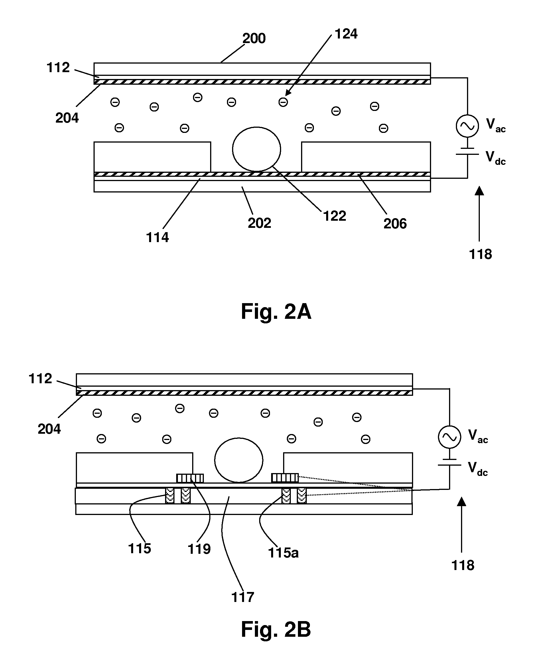
[0095]Referring now to FIG. 4, a prototype device for showing electric field directed movement of charged molecules is illustrated. The device comprises a sheet of a 250 μM thick silicone gasket material 410 having a 1 mm diameter hole in the sheet. This is applied to a 20 nm parylene dielectric layer 408 which had been applied to a 150 nm ITO layer 406 on a 1 in.×3 in glass slide 404, forming a “bottom.” To form a “top,” another glass slide 414 was similarly coated with an ITO layer 415 to form an electrode and a parylene layer 417 on the “bottom” side of the top electrode in contact with fluid, which can be contained in a well 412 formed when the two slides were sandwiched together.

[0096]A Coolsnap fx-16 CCD camera, Olympus IX70 epifluorescent microscope imaging device 416 was arrayed beneath the fluid well. Conductive copper tape was used to connect the electrodes to the signal generator.

[0097]10 mM Tris-HCl solution ...

example 3

Electric Field Directed Preconcentration of Pyrophosphate

[0099]A device was constructed essentially as shown in FIG. 4, with the following differences: instead of a camera and microscope, a magnet and a Hamamatsu photomultiplier tube was arrayed beneath the well 412, as shown at 416 in FIG. 4.

[0100]The procedure was as follows: Conductive copper tape was attached to the exposed ITO layer on the glass slides. The silicone gasket was placed on the bottom glass slide and pressed against the slide to form a seal. The 1 mm hole in the gasket was loaded with magnetic beads containing enzymes. A magnet was placed below the glass slide to hold the magnetic beads stationary. The 1 mm hole in the gasket was filled with pyrophosphate solution. The second glass slide was placed on the top of the gasket to seal the top of the well containing the chemicals. The chemicals were as follows: Pyrophosphate solution, magnetic beads loaded with ATP sulfurylase, and luciferase (obtained from 454 Life Sci...


PUM

PropertyMeasurementUnit
Thicknessaaaaaaaaaa
Electric potential / voltageaaaaaaaaaa
Frequencyaaaaaaaaaa

Abstract

Disclosed are a method and apparatus that use an electric field for improved biological assays. The electric field is applied across a device having wells, which receive reactants, which carry a charge. The device thus uses a controllable voltage source between the first and second electrodes, which is controllable to provide a positive charge and a negative charge to a given electrode. By controlled use of the electric field charged species in a fluid in a fluid channel are directed into or out of the well by an electric field between the electrodes. The present method involves the transport of fluids, as in a microfluidic device, and the electric field-induced movement of reactive species according to various assay procedures, such as DNA sequencing, synthesis or the like.

Description

CROSS-REFERENCE TO RELATED APPLICATIONS[0001]This application claims priority from U.S. Provisional Patent Application No. 60 / 959,398 filed on Jul. 13, 2007, which is hereby incorporated by reference in its entirety.STATEMENT OF GOVERNMENTAL SUPPORT[0002]None.REFERENCE TO SEQUENCE LISTING, COMPUTER PROGRAM, OR COMPACT DISK[0003]NoneBACKGROUND OF THE INVENTION[0004]1. Field of the Invention[0005]The present invention relates to the field of biological assays and apparatus for carrying out such assays, such as a microfluidic device, to which is applied electric fields to control movement of charged molecules. The assays involve charged molecular species, such as nucleotides (due to phosphate ions), or other molecules which contain a charge due to their ionic nature, such as certain proteins or small molecules.[0006]2. Related Art[0007]Advances in silicon microfabrication have been used to produce microchannels and microarrays for many lab-on-a-chip platforms. Advantages include low re...

Claims


Application Information

Patent Timeline
IPC IPC(8): B01D59/42
CPCB01J2219/00317B01J2219/00459B01J2219/00466B01J2219/00596B01J2219/00648B01L2400/0415B01J2219/00702B01L3/502761B01L2200/0668B01L2300/0819B01J2219/00653
Inventor RONAGHI, MOSTAFAKHURANA, TARUNSANTIAGO, JUAN G.
Owner THE BOARD OF TRUSTEES OF THE LELAND STANFORD JUNIOR UNIV
Why Eureka
Try Eureka