Spine three-dimensional positioning method

A positioning method and positioning algorithm technology, applied in the direction of stereotaxic surgical instruments, puncture needles, trocars, etc.

Active Publication Date: 2018-08-28
BEIJING HOSPITAL
View PDF3 Cites 2 Cited by
  • Summary
  • Abstract
  • Description
  • Claims
  • Application Information

AI Technical Summary

Problems solved by technology

However, there is currently a lack of a simple and accurate ...

Method used


Image

  • Spine three-dimensional positioning method
  • Spine three-dimensional positioning method
  • Spine three-dimensional positioning method

Examples


Embodiment 1

[0114] Embodiment 1. Positioning method for spinal puncture based on three-dimensional space positioning algorithm

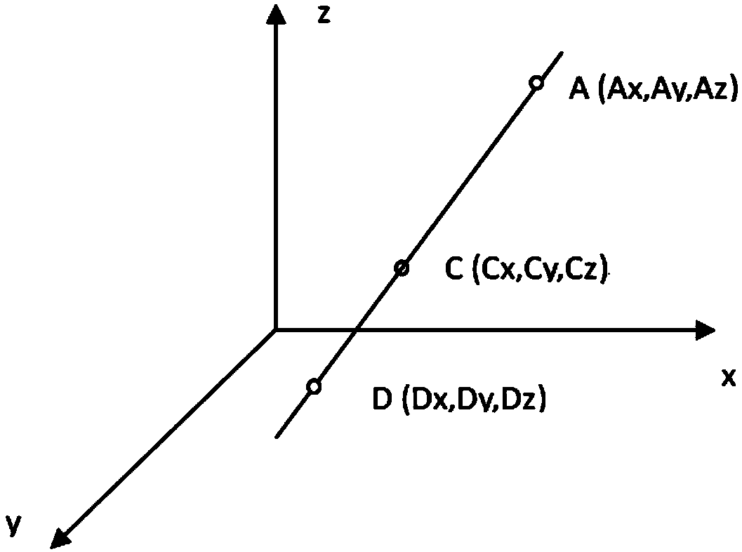
[0115] Step 1, obtain the spine front view through the X-ray photograph ( Figure 4 ) and profile ( Figure 5 ) image, set the target bit reference scale of the front bitmap and the side bitmap when taking pictures;

[0116] Step 2, import the X-ray photo image, and determine the spatial positions of the marker points C and D in the front view and the lateral view according to the given scale;

[0117] Step 3, respectively mark the origin and the coordinate system in the images of the front view and the side view, and set the scale;

[0118] Step 4. Mark the relevant points in the image: the longitudinal coordinates of the puncture start point A, the skin entry point B and the puncture end point E, and the system calculates and generates the coordinate values ​​of each point;

[0119] (1) Bring the known Az coordinates into the equation to get the Ax and Ay v...

Embodiment 2

[0149] Embodiment 2. Develop computing system with the method of the present invention

[0150] 1. System composition and calculation method

[0151] refer to Figure 7 As shown, the system includes the following modules: image input module (B1), used to import X-ray photos; image analysis module (B2), used to set the coordinate system of the image, set the scale of the image, and mark the relevant points on the image The data checking module (B3) is used to check the integrity of the data; the data calculation and display module (B4) is used to calculate and output the calculation results of the puncture position and angle.

[0152] The image input by the image input module is an X-ray photo, including an anteroposterior image of the spine and a lateral image of the spine; the image analysis module has an image angle calibration function.

[0153] The relevant points are the longitudinal coordinates of the puncture start point A, the skin entry point B, the puncture end poi...

Embodiment 3

[0192] Embodiment 3. method verification experiment

[0193] 1. Materials and methods

[0194] 1. Materials:

[0195] Select 4 adult spine cadavers with complete T2-S1 dorsal structure (skin and soft tissue) to verify the puncture instrument and calculation method. Each specimen can provide a total of 12 bilateral L1-S1 pedicle puncture locations, and a total of 48 punctures were performed.

[0196] Before the puncture, the specimens underwent spinal computerized tomography (CT) and X-ray examination.

[0197] 2. Method:

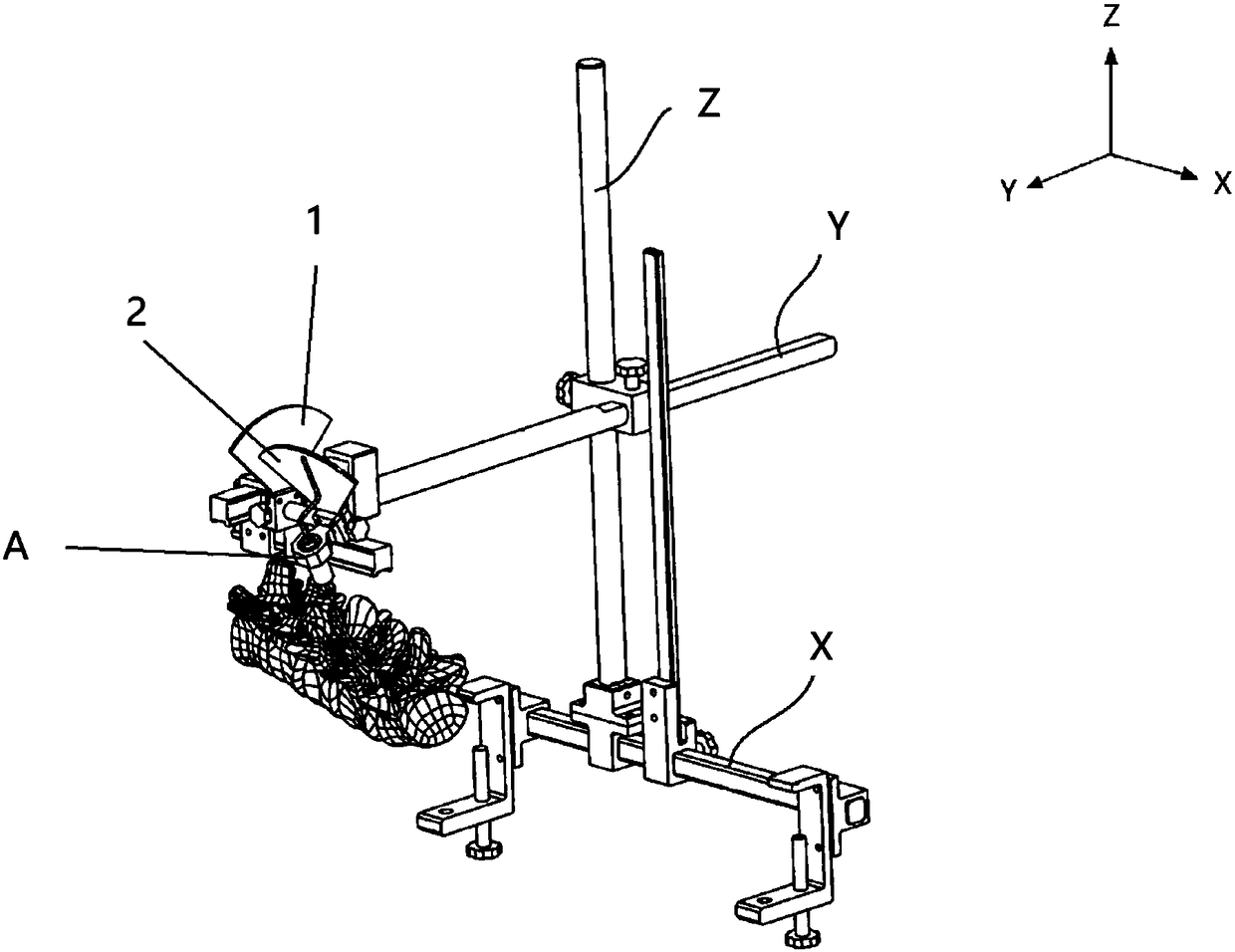
[0198] Puncture process and locator ( figure 1 and figure 2 ), calculation software (embodiment 2) method of use is the same as the preceding description. Input the revised standard coordinate values ​​into the calculation software to obtain the coordinates of the puncture starting point and the puncture direction, adjust the parameters of the locator, and start the puncture. The puncture needle used in this experiment was a Kirschner wire. When th...


PUM


Abstract

The invention discloses a spine three-dimensional positioning method and relates to the technical field of minimally invasive surgery. The method includes: acquiring a normal position image and a sideposition image of a spine through X-ray photography; guiding in an X-ray photography image, and determining spatial positions of mark points C and D in each of the normal position image and the sideposition image according to a given scale; respectively marking original point and coordinate system in the normal position image and the side position image; marking related points in the images, andcalculating to generate coordinate values of each point by a system; determining a spatial straight line according to two points, calculating spatial coordinates of the related points and included angles between the straight line determined by the points C and D and a horizontal plane and a vertical plane, and determining puncture position, angle and distance of a puncture needle through a triangular function; according to output parameters, setting position and angle of the puncture needle of a spine puncture locator. By the method, complex calculation process is made quite simple and convenient, and visual puncture line prompting is combined, so that errors caused by calculation are avoided basically.

Description

technical field [0001] The invention discloses a three-dimensional spine positioning method, which relates to the technical field of minimally invasive surgery, in particular to a spine minimally invasive positioning system and its application in spinal minimally invasive positioning. Background technique [0002] Minimally invasive surgery is the application of advanced tools (such as computers, special puncture guide needles, special retractors and imaging equipment) to complete traditional operations through special surgical approaches, so as to achieve the least tissue damage to patients, the lightest The psychological impact, the fastest recovery and the best surgical results. [0003] Minimally invasive spinal surgery is a surgical method for the treatment of various spinal diseases (such as osteoporotic compression fractures, spinal traumatic fractures, lumbar disc herniation, lumbar spinal stenosis, etc.) that has emerged in recent years. Small size, short operation...

Claims


Application Information

Patent Timeline
IPC IPC(8): A61B90/11A61B17/34
CPCA61B17/3403A61B17/3472A61B17/3478A61B90/11
Inventor 王作伟
Owner BEIJING HOSPITAL
Why Eureka
Try Eureka