Mouse fibroblast and application thereof
A technology of fibroblasts and mice, applied in the field of cell biology, can solve the problems of cross-contamination, misidentification of mammalian cells, etc.
- Summary
- Abstract
- Description
- Claims
- Application Information
AI Technical Summary
Problems solved by technology
Method used
Image
Examples
Embodiment 1
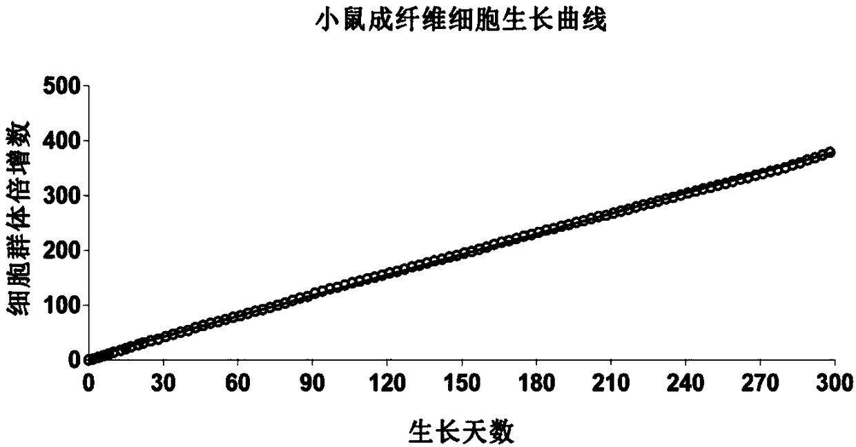
[0100] [Example 1] Primary isolation culture and subculture of mouse fibroblasts
[0101] Including the following steps:
[0102] (1) 16-18 day old Swiss embryonic mice were taken, and normal embryonic dorsal skin tissue was collected.
[0103] (2) Wash the isolated tissue samples with 95-100% (v / v) ethanol, then wash 3 times with PBS (0.01M, pH7.4), and then put the tissue samples into sterile culture medium containing pre-cooled PBS In a dish, mince the tissue with dissecting forceps and scissors.
[0104] (3) Digest the chopped tissue sample with digestive juice; preferably, the digestive juice is HL culture medium containing collagenase and dispase.
[0105] (4) The digested tissue was centrifuged at 1000 rpm for 5 minutes to remove the supernatant, and the cell pellet was resuspended in 0.25% (mass volume ratio) trypsin-EDTA for digestion.
[0106] (5) Add DMEM medium containing 10% (v / v) FBS, and centrifuge at 1000 rpm for 5 minutes to remove the supernatant.
[0107...
Embodiment 2
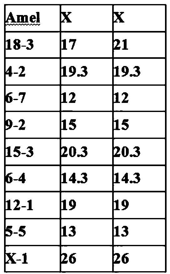
[0113] [Example 2] Genotyping analysis and identification of mouse fibroblasts
[0114] Including the following steps:
[0115] (1) Mouse fibroblasts (1×10 6 ), wash the cells twice with 1×PBS, digest the monolayer cells with 0.05% trypsin-EDTA for 30s, and neutralize the digestion reaction with 10 mL of complete DMEM;
[0116] (2) Centrifuge at 10,000 rpm for 1 min, pour off the supernatant, add 200 μL buffer GA (cell / tissue genomic DNA extraction kit DP304, Tiangen Company), and shake until completely suspended;
[0117] (3) Add 20 μL Proteinase K solution and mix well;
[0118] (4) Add 200 μL of buffer solution GB (cell / tissue genomic DNA extraction kit DP304, Tiangen Company), fully invert and mix well, place at 70°C for 10 min, and briefly centrifuge;
[0119] (5) Add 200 μL of absolute ethanol, shake and mix well for 15 seconds, and briefly centrifuge;
[0120] (6) Add the resulting solution and the flocculent precipitate into an adsorption column (cell / tissue genomi...
Embodiment 3
[0128] [Example 3] Treating mouse fibroblasts with the drug Mitomycin C to make them lose their ability to proliferate
[0129] Including the following steps:
[0130] (1) When the mouse fibroblasts grow to 80-90%, add mitogenin C (dissolved in water, stock solution concentration: 0.5 mg / mL) with a final concentration of 10 μg / mL in the culture medium, and store at 37 °C Processing 2h;
[0131] (2) Then add 1xPBS or serum-free medium (DMEM) in warm bath to wash 3 times, and discard the washing solution;
[0132] (3) Add 0.05% trypsin / EDTA to predigest the cells for 30-40s, then discard them, add 0.05% trypsin / EDTA to digest them for 30s, tap the culture dish to disperse the cells, and then add complete medium (containing 10% fetal calf DMEM) neutralization reaction of serum;
[0133] (4) Low-speed centrifugation (1000rpm) to remove the supernatant and obtain cell pellets;
[0134] (5) Although the precipitated cells lose their proliferative ability and still maintain metabol...
PUM
 Abstract
Description
Claims
Application Information
 - R&D
- Intellectual Property
- Life Sciences
- Materials
- Tech Scout
- Unparalleled Data Quality
- Higher Quality Content
- 60% Fewer Hallucinations
Browse by: Latest US Patents, China's latest patents, Technical Efficacy Thesaurus, Application Domain, Technology Topic, Popular Technical Reports.
© 2025 PatSnap. All rights reserved.Legal|Privacy policy|Modern Slavery Act Transparency Statement|Sitemap|About US| Contact US: help@patsnap.com


