

Thyroid tumor ultrasonic image recognition method and device
A technology for thyroid tumors and ultrasound images, applied in the field of image recognition, can solve problems such as low recognition accuracy
- Summary
- Abstract
- Description
- Claims
- Application Information
AI Technical Summary
Problems solved by technology
Method used
Image

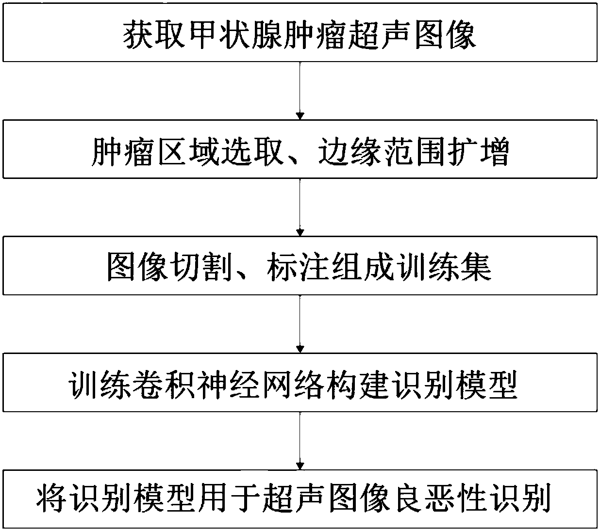
Examples
Embodiment 1
[0073] Example 1 Obtaining Ultrasonic Images of Thyroid Tumors
[0074] The original dataset was collected from patients with thyroid nodules by the Cancer Hospital of Fudan University from January 2014 to December 2016. All papillary thyroid carcinoma (PTC) lesions were pathologically confirmed by surgical resection specimens. Each image in the raw data set was ranked by three experienced radiologists using the Thyroid Imaging Reporting and Data System (TI-RADS). TI-RADS scores are divided into 2, 3, 4a, 4b, and 4c, respectively: no suspicion, probably benign nodules, a single suspicious feature, two suspicious features, and three or more suspicious features. Three radiologists also acquired sonographic features, including their composition, echogenicity, calcifications, margins, and shape.
[0075] The entire dataset has a total of 2836 original images, of which 1484 are PTC-diagnosed ultrasound images and 1352 are collected from benign tumors, including nodular diseases a...
Embodiment 2
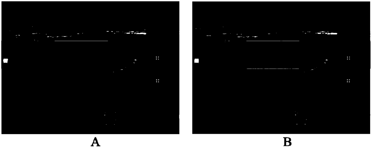
[0076] Example 2 Tumor Region Selection, Margin Range Expansion
[0077] All images in the original data set are collected from the phenomenon reporting system. If the entire image is used as a training sample, its inherent background, text, etc. will greatly affect the extraction of benign and malignant features of tumors. Therefore, it is necessary to select the tumor area on the original image, that is, to use a rectangular frame to completely select the tumor block, such as figure 2 shown in the inner rectangle box in . The selection process is completed one by one by doctors using semi-automated selection scripts.
[0078] figure 2 The area shown in the middle and inner rectangular frame only includes the nodule itself, and the information of the surrounding tissue of the nodule is very necessary for the judgment of benign and malignant tumors, so the concept of margin range is added when cutting the image. figure 2 The outer rectangular frame in the two images is t...
Embodiment 3
[0082] Embodiment 3 image cutting, labeling form training set
[0083] The original data set was split according to the ratio of 6:1. The original data set has a total of 2836 images. In the experiment, 1275 PTC images and 1162 benign images were randomly selected as the training set, and the remaining 209 PTC images and 190 benign images were used as the test set. In the process of data set segmentation, it is noted that the images of the same patient can only exist in the training set or the test set at the same time, which prevents the interference of similar images.
[0084] In the process of experimental testing, the size of the tumor itself is also considered to be quite different, so the data set is divided into smaller than 0.5cm, 0.5cm to 1cm, and larger than 1cm. The distribution on Figure 4 As shown, the same distribution of training and testing data is satisfied.
PUM
 Abstract
Description
Claims
Application Information