Ureteroscope of separated structure

A ureteroscope and split-type technology, applied in the field of ureteroscope, can solve the problems that ordinary medical staff cannot disassemble without permission, the specifications and models are limited, and the unit price of ureteroscope is high, so as to prevent medical accidents of cross-infection and effectively utilize resources. High, improve the effect of surgical safety

Pending Publication Date: 2018-10-09
江门市硕通医疗器械科技有限公司
View PDF9 Cites 1 Cited by
  • Summary
  • Abstract
  • Description
  • Claims
  • Application Information

AI Technical Summary

Problems solved by technology

[0003] At present, the ureteroscopes produced by domestic and foreign medical device manufacturers are all highly integrated. For example, the endoscope tube, the optical lens and the image beam in it are glued or welded together by optical glue, and ordinary medical staff cannot disassemble it without permission. However, this highly integrated structure has the following problems in use:
[0004] (1) The damaged ureteral endoscope needs to be returned to the factory as a whole, and can only be repaired after disassembly by professional personnel through technical means, which will affect the effective use
[0005] (2) Re-cleaning, disinfection and sterilization are required after each operation. Due to the repeated use of instruments, disinfection and sterilization work is difficult to guarantee, and there are medical risks of cross-infection of infectious diseases
[0006] (3) Due to the high unit price of ureteral endoscopes, it is usually divided into three specifications: 8 / 9.8Fr, 6 / 7.5Fr, and 4.5 / 6Fr according to the thickness of the endoscope catheter and the size of the objective lens end. The model is limited, and it cannot meet the requirements for some special patients such as adult patients with relatively narrow ureters, newborns or young children

Method used


Image

  • Ureteroscope of separated structure
  • Ureteroscope of separated structure
  • Ureteroscope of separated structure

Examples


Embodiment Construction

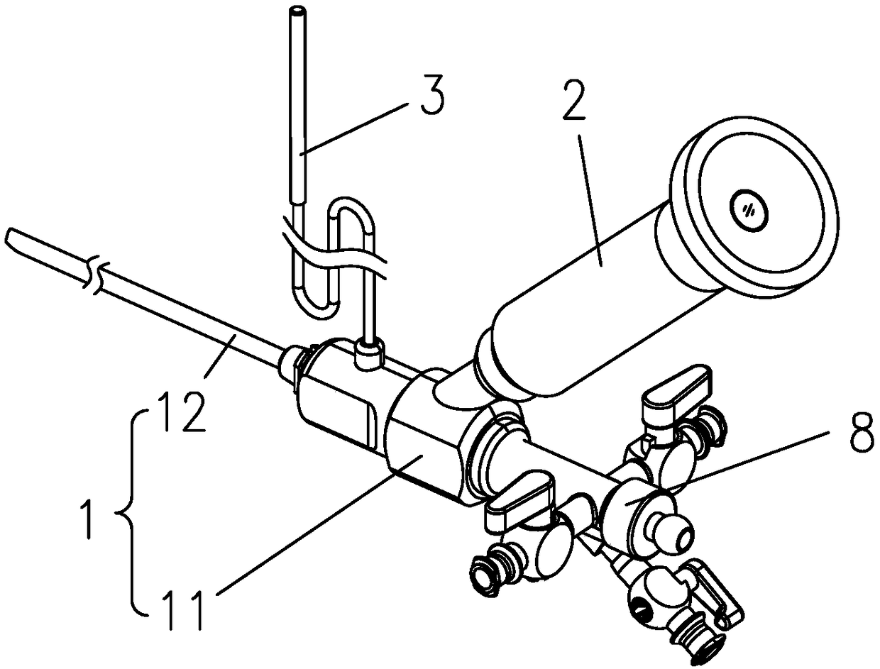
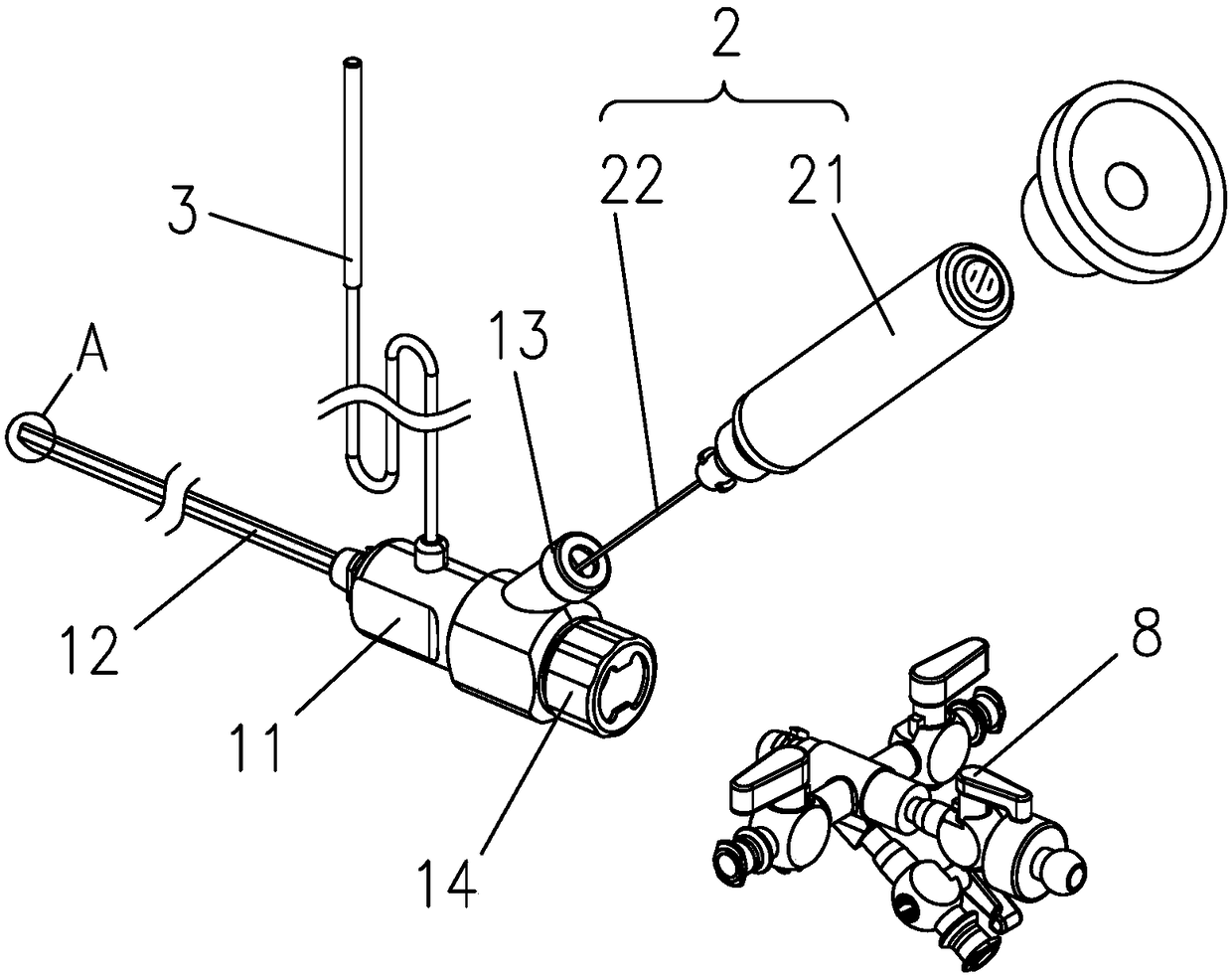
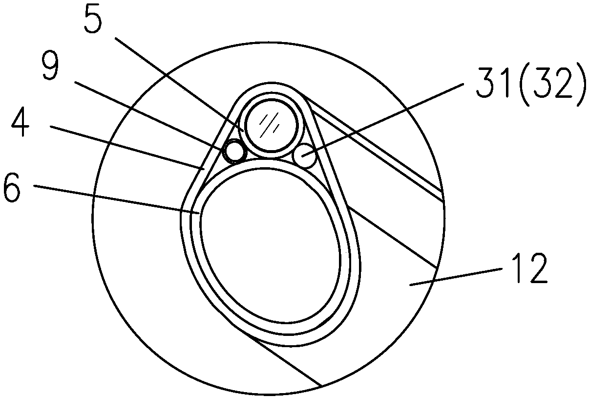
[0027] refer to Figure 1 to Figure 4 , a ureteroscope with a split structure, including a mirror body 1 and an image transmission assembly 2 and an illumination assembly 3 provided thereon; the mirror body 1 includes a handle 11 and a working pipeline 12 protruding from the front end of the handle 11 . Catheter 12 can be rigid or flexible catheter, and it comprises outer mirror tube 4 and the endoscope tube 5 and working pipeline 6 that are positioned at the outer mirror tube 4 pipe body, and image transmission assembly 2 then includes imaging device 21 and puts endoscope tube Image beam 22 in 5.

[0028] Wherein, the mouth of the endoscope tube 5 is sealed and covered with a flat lens 51 to form a sealed end, effectively ensuring that the image transmission bundle 22 etc. during the period will not come into contact with the human body; and between the image transmission assembly 2 and the mirror body 1 is A detachable installation structure, and an optical lens 7 is integr...


PUM


Abstract

The invention discloses a ureteroscope of a separated structure. The ureteroscope comprises a ureteroscope body and further comprises an image transmitting assembly and an illuminating assembly whichare arranged on the ureteroscope body. The ureteroscope body comprises a handle part and a catheter stretching out of the front end of the handle part; the catheter comprises an outer ureteroscope tube and further comprises an inner ureteroscope tube and a working pipeline which are arranged in the outer ureteroscope tube; the image transmitting assembly comprises an imaging device and an image transmitting bundle arranged in the inner ureteroscope tube, a covering lens is arranged at a tube opening of the inner ureteroscope tube in a sealing covering mode, and thus a sealed end is formed; anoptical lens is integrated at the front end of the image transmitting bundle; and the image transmitting assembly is detachably mounted on the mirror body so that when the imaging device is detached,the image transmitting bundle on the imaging device can be pulled away from the inner ureteroscope tube synchronously. According to the separated type ureteroscope, the equipping and using costs of the ureteroscope can be lowered, the using efficiency is improved, meanwhile, the existing modern mode is improved, and the more effective, more hygienic and safer effects are achieved.

Description

technical field [0001] The invention relates to a medical device, in particular to a ureteroscope with a split structure. Background technique [0002] Ureteroscopes are mainly used in upper urinary tract microscopy, and can be used for biopsy, lithotripsy, stone removal and other operations with other surgical instruments. The catheter protruding from the front end of the handle, the catheter includes the outer mirror tube and the inner mirror tube and working pipeline; the image transmission component includes the imaging device and the image transmission bundle placed in the endoscope tube, and the image transmission component is the core of the whole device , occupies the main cost of the endoscope. [0003] At present, the ureteroscopes produced by domestic and foreign medical device manufacturers are all highly integrated. For example, the endoscope tube, the optical lens and the image beam in it are glued or welded together by optical glue, and ordinary medical staff...

Claims


Application Information

Patent Timeline
Patent Type & Authority Applications(China)
IPC IPC(8): A61B1/307A61B1/04
CPCA61B1/00064A61B1/00112A61B1/04A61B1/307
Inventor 周均洪
Owner 江门市硕通医疗器械科技有限公司
Why Eureka
Try Eureka