Method for establishing body lumbar three-dimensional simulation model through registration and fusion of CT (Computed Tomography) and MRI (Magnetic Resonance Imaging) signals

A 3D model and 3D simulation technology, applied in the field of medical imaging virtual simulation, can solve problems such as inability to obtain common registration points of bone tissue

Active Publication Date: 2018-10-26
HUAQIAO UNIVERSITY
View PDF9 Cites 23 Cited by
  • Summary
  • Abstract
  • Description
  • Claims
  • Application Information

AI Technical Summary

Problems solved by technology

The proportion of bone tissue in the lumbar spine and the knee joint is different, and this method cannot obtain better bone tissue as a common registration point for the two images

Method used


Image

  • Method for establishing body lumbar three-dimensional simulation model through registration and fusion of CT (Computed Tomography) and MRI (Magnetic Resonance Imaging) signals
  • Method for establishing body lumbar three-dimensional simulation model through registration and fusion of CT (Computed Tomography) and MRI (Magnetic Resonance Imaging) signals
  • Method for establishing body lumbar three-dimensional simulation model through registration and fusion of CT (Computed Tomography) and MRI (Magnetic Resonance Imaging) signals

Examples


Embodiment 1

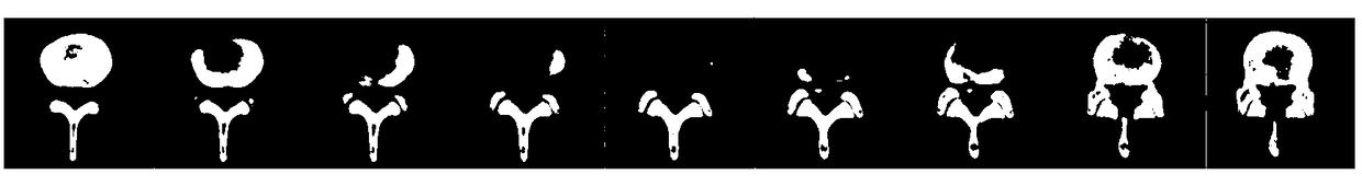
[0149] Two sets of CT and MRI images of "fourth lumbar vertebrae / fifth lumbar vertebral right lateral recess stenosis combined with fifth lumbar upper endplate vertebral posterior edge disconnection" were used to construct a 3D model, and then registered and fused into a 3D model Designing the surgical approach for percutaneous transforaminal discectomy.

[0150] In this embodiment, in the three-dimensional model of the lumbar spine that is registered and constructed by the method of the present invention, the three-dimensional model of the lumbar intervertebral disc, dural sac and nerve root is consistent with the three-dimensional model of the lumbar intervertebral disc, dural sac and nerve root established using computer tomography data. It has the advantages of accurate spatial position, clear and smooth outline, and clear adjacency relationship.

[0151] see Figure 1-1 to Figure 1-21 As shown, the present embodiment a kind of method for registration fusion CT and MRI si...

Embodiment 2

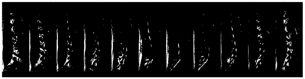
[0273] Two sets of CT and MRI two-dimensional images of "third lumbar vertebra / fourth lumbar disc herniation combined with third lumbar lower endplate vertebral body disconnection" were used to construct a three-dimensional model, and then registered and fused into a three-dimensional model. Design the surgical approach, 3D print the physical model, and compare and analyze the preoperative and postoperative results of transforaminal endoscopic discectomy.

[0274] In this embodiment, the three-dimensional model of the lumbar intervertebral disc registered and constructed by this method has a higher spatial resolution than the lumbar intervertebral disc model established based on the routine examination sequence (T1WI sequence, T2WI sequence, T2FS sequence) data of magnetic resonance imaging. It has the advantages of accurate spatial position, clear and smooth outline, and clear adjacency relationship.

[0275] see Figure 2-1 to Figure 2-25 As shown, a method of registering a...


PUM


Abstract

The invention relates to a method for establishing a body lumbar three-dimensional simulation model through registration and fusion of CT (Computed Tomography) and MRI (Magnetic Resonance Imaging) signals. The method comprises the steps of collecting a computed tomography (CT) image; collecting a magnetic resonance imaging (MRI) image; establishing a computed tomography image three-dimensional model; establishing a magnetic resonance imaging image three-dimensional; and carrying out registration and fusion on the computed tomography image three-dimensional model and the magnetic resonance imaging image three-dimensional, wherein the registration and fusion comprise carrying out simple registration and global computing registration according to a lumbar anatomic structure. According to themethod, existing routine examinations such as computed tomography and magnetic resonance imaging are utilized fully, through combination of optimized magnetic resonance scanning sequences, lumbar intervertibral disc, nerve root and ligamentum flavum important soft tissue three-dimensional models capable of mutually verifying accuracy in various magnetic resonance sequences are established, a new lumbar intervertibral disc medical image high-accuracy modeling mode is established, and a utilization rate of medical image examination data is greatly improved.

Description

technical field [0001] The invention belongs to the field of virtual simulation of medical images, and in particular relates to a method for using two-dimensional data of computerized tomography and magnetic resonance imaging to respectively establish three-dimensional models and then register and fuse them to form a high-resolution virtual simulation three-dimensional model, which is suitable for human lumbar spine Auxiliary diagnosis of organic lesions and making surgical decisions. Background technique [0002] Orthopedic patients usually need to undergo necessary imaging examinations before surgery, including CT (Computed Tomography, computerized tomography) and MRI (Magnetic Resonance Imaging, magnetic resonance imaging). CT can accurately detect the slight difference in density between different tissues on a transverse anatomical plane, and is an ideal examination method for observing bone, joint and soft tissue lesions. MRI has a wide range of tissue density contrast...

Claims


Application Information

Patent Timeline
Patent Type & Authority Applications(China)
IPC IPC(8): G06T17/00G06T7/33G16H30/20
CPCG06T7/33G06T17/00G16H30/20G06T2207/20221
Inventor 郑志发林俊生
Owner HUAQIAO UNIVERSITY
Why Eureka
Try Eureka